Man is a complex and subtle substance responsible for the regulation of the whole organism.
The structure and features of the functioning of this organ are still not fully understood, scientists every day discover new features that allow the brain to work. But such a part as the black substance of the brain or substantia nigra has long been known.
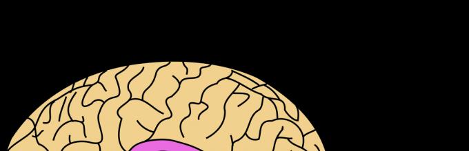
What is black substance and where is it located?
The substance is one of the oldest parts in the structure of the brain, located in its core - the quadrigemina of the midbrain. Historically, she was responsible for the movements of our ancestors, then, when they became more complicated, the structure also changed. The black substance was overgrown with nerve connections, forming a more complex structure.
The black substance got its name due to the action of the pigment - neuromelanin, which stains the cells in a dark color. The black matter of the midbrain is heterogeneous, it is divided into two halves: right and left. In addition, two layers are distinguished in the substance: ventral and compact. The ventral is closer to the front of the head, and the compact is to the back. The first provides the synthesis of the neurotransmitter, the second is engaged in the processing of incoming information and its transfer to other structures.
Ultrasound of the substantia nigra of the brain shows that it is connected with all departments, but most closely with the basal ganglia and visual tubercles.

An abundant supply of blood indicates the high role of structure in the activity of the body. Among the functions of the black substance are:
- implementation of elementary movements: swallowing, chewing, breathing, eye movements and others;
- regulation of small and precise movements of the limbs;
- help in expressing emotions;
- participation in emotional processes;
- causing some mental disorders.
The role of the substance in the development of pathologies
The role of substance in the development of mental illness is great. It contains, the processes of which diverge throughout the brain, affecting the legs of the brain and the internal capsule.
Their endings, containing terminal microvesicles that produce dopamine, are located in. Any damage to this structure leads to disturbances in motor functions and psycho-emotional.
Schizophrenia
The mechanism of development has been studied for many years, but researchers have not come to a consensus. are being put forward different theories formation of this disease, one of which links schizophrenia with disorders in the work of the substantia nigra, the nucleus of which is located in the midbrain. This is the so-called dopamine hypothesis.
Studies show that patients with schizophrenia have deviations in the synthesis and perception of dopamine.
So, they show:
- increased production of the hormone;
- an increase in the concentration of dopamine in synapses;
- increased production;
- more dopamine released when exposed to amphetamines.
Such hypersensitivity contributes to excessive stimulation of brain neurons and its overexcitation. The patient is unable to control the stream of consciousness, and his perception of the surrounding reality changes. Similar conditions occur when taking psychotropic substances, which in healthy people cause hallucinations and other abnormalities, but in sick people they act much more strongly.
According to statistics, the development of schizophrenia captures more men than women. In the former, it usually develops earlier and is more severe. For women, the onset of symptoms at the age of 25-30 years is typical.
The substantia nigra made it possible to examine its structure and discover that in schizophrenia, changes in the dopamine system affect the associative striatum more than the limbic one.
Moreover, deviations in the synthesis of dopamine by neurons in the direction of increasing its production are observed even before the development of the disease. But the more likely to get schizophrenic, the higher these deviations.
The reasons for their appearance, scientists call several points:
- disturbances in the control system of the influence of the hippocampus on the dopamine pathway;
- changes occurring in the structure of neurons that produce a neurotransmitter;
- malfunctions in the work of cortical structures that affect dopamine systems;
- influence of other neurotransmitter systems.
Thus, changes in the work of the dopamine system are observed in all cases of schizophrenia. However, the influence of other brain structures is not excluded.
Treatment based on blocking receptors or inhibition of dopamine production in most cases gives positive results. Patients are offered antipsychotic drugs that block the action of dopamine, although they have a serious consequence - depressive states. Safer is cognitive-behavioral therapy, which can be carried out by a psychologist.
Parkinson's disease
When there is damage to dopaminergic neurons located in the compact part of the substantia nigra, in which aggregation occurs throughout the brain.
In this case, strong motor, psychopathological and cognitive anomalies arise:
- slowdown;
- decrease or absence of facial expressions;
- tremor;
- the predominance of the flexion posture;
- the complexity of the transition from a state of rest to a state of motion;
- memory impairment;
- and others.
The disease develops mainly in men over the age of 60, women suffer from this pathology less often. The reason is changes in the dopamine system, which can be caused both by poisoning with toxic substances and by taking certain drugs. In addition, the disease develops without "apparent" causes, the source of which has not yet been discovered. Many facts indicate a hereditary predisposition to Parkinson's disease.
Scientists suggest that the destruction of neurons begins in the peripheral nervous system, and then goes to the brain stem and moves to the middle and anterior. Support for this theory is found in the change in cholinergic neurotransmission seen in patients with mild disease.
With the defeat of 30 percent of the neurons that produce dopamine, its deficiency occurs and the symptoms of the disease develop. Its distribution occurs unevenly and usually progresses in the direction from the back of the black substance to the front.
The studies also found MPTP neurotoxin, which destroys dopamine neurons. It is important to understand the source of its products and try to limit it.
The use of therapy with the use of L-dopa, an intermediate element in the dopamine production scheme, gives a positive result in terms of disease inhibition. However, it does not allow restoring the lost brain structures. In addition, with the progression of Parkinson's disease, the effectiveness of this therapy is markedly reduced.
Consequences of damage
Neurons in the substantia nigra of the midbrain produce neurotransmitters, the main of which is dopamine. It acts as a means of "rewarding" the brain, causing feelings of pleasure and influencing the motivation and learning of a person.
Yes, application psychotropic drugs or drug causes a big surge of dopamine and, as a result, pleasure. In an attempt to repeat it, the person begins to use the stimulant regularly. However, such surges are compensated nervous system and so-called tolerance is developed - reduced sensitivity to the action of a substance. As a result, the level of pleasure gradually decreases, but the desire to receive it remains.
This and other features of the action chemical substances, including drugs, are being investigated in neuropharmacology and toxicology.
The components under study include:
- cocaine;
- amphetamines;
- MPTP;
- Levodop.
Cocaine and amphetamines are dopamine-enhancing and addictive substances. In addition, they can "push" the development of schizophrenia.
The latter is used in the treatment of Parkinson's disease, is quite effective in eliminating symptoms, but does not work in restoring lost structures.
MPTP stands for methylphenyltetrahydropyridine and is a neurotoxin that directly reduces dopamine production. Now used by scientists to model the disease in animals in an attempt to understand the mechanism of its development.
On its ventral surface there are two massive bundles of nerve fibers - the legs of the brain, through which signals are carried from the cortex to the underlying structures of the brain.
Rice. 1. The most important structural formations of the midbrain (cross section)
In the midbrain, there are various structural formations: the quadrigemina, the red nucleus, the substantia nigra, and the nuclei of the oculomotor and trochlear nerves. Each formation performs a specific role and contributes to the regulation of a number of adaptive reactions. All ascending paths pass through the midbrain, transmitting impulses to the thalamus, cerebral hemispheres and cerebellum, and descending paths, conducting impulses to the medulla oblongata and spinal cord. The neurons of the midbrain receive impulses through the spinal and medulla oblongata from the muscles, visual and auditory receptors along the afferent nerves.
Anterior colliculi are the primary visual centers, and they receive information from the visual receptors. With the participation of the anterior tubercles, visual orienting and watchdog reflexes are carried out by moving the eyes and turning the head in the direction of the action of visual stimuli. The neurons of the posterior tubercles of the quadrigemina form the primary auditory centers and, upon receiving excitation from the auditory receptors, ensure the implementation of auditory orienting and sentinel reflexes (the animal's auricles tense up, it becomes alert and turns its head towards a new sound). The nuclei of the posterior tubercles of the quadrigemina provide a sentinel adaptive reaction to a new sound stimulus: redistribution of muscle tone, increased tone of the flexors, increased heart and breathing contractions, increased blood pressure, i.e. the animal prepares for defense, flight, attack.
black substance receives information from muscle receptors and tactile receptors. It is associated with the striatum and the globus pallidus. Neurons of the substantia nigra are involved in the formation of an action program that coordinates the complex acts of chewing, swallowing, as well as muscle tone and motor reactions.
red core receives impulses from muscle receptors, from the cerebral cortex, subcortical nuclei and cerebellum. Has a regulatory effect on motor neurons spinal cord through the nucleus of Deiters and the rubrospinal tract. The neurons of the red nucleus have numerous connections with the reticular formation of the brain stem and, together with it, regulate muscle tone. The red nucleus has an inhibitory effect on the extensor muscles and an activating effect on the flexor muscles.
Eliminating the connection of the red nucleus with the reticular formation of the upper part of the medulla oblongata causes a sharp increase in the tone of the extensor muscles. This phenomenon is called decerebrate rigidity.

|
Name |
midbrain functions |
|
Kernels of the roof of the superior and inferior tubercles of the quadrigemina |
Subcortical centers of vision and hearing, from which the tectospinal path originates, through which orienting auditory and visual reflexes are carried out |
|
The nucleus of the longitudinal medial bundle |
Participates in providing a combined turn of the head and eyes to the action of unexpected visual stimuli, as well as irritation of the vestibular apparatus |
|
Nuclei III and IV pairs of cranial nerves |
They participate in the combination of eye movement due to the innervation of the external muscles of the eye, and the fibers of the autonomic nuclei after switching in the ciliary ganglion innervate the muscle that narrows the pupil and the muscle of the ciliary body |
|
Red cores |
They are the central link of the extrapyramidal system, since the paths from the cerebellum (tr. cerebellotegmenlalis) and the basal nuclei (tr. pallidorubralis) end on them, and the rubrospinal path begins from these nuclei |
|
black substance |
It has a connection with the striatum and the cortex, participates in complex coordination of movements, regulation of muscle tone and posture, as well as in coordinating the acts of chewing and swallowing, is part of the extrapyramidal system |
|
Kernels of the reticular formation |
Activating and inhibitory effects on the nuclei of the spinal cord and various areas of the cerebral cortex |
|
Gray central periaqueductal substance |
Part of the antinociceptive system |
The structures of the midbrain are directly involved in the integration of heterogeneous signals necessary for the coordination of movements. With the direct participation of the red nucleus, the black substance of the midbrain, the neural network of the stem movement generator and, in particular, the eye movement generator, is formed.
Based on the analysis of signals entering the stem structures from proprioreceptors, vestibular, auditory, visual, tactile, pain and other sensory systems, a flow of efferent motor commands is formed in the stem movement generator, sent to the spinal cord along descending pathways: rubrospinal, retculospinal, vestibulospinal, tectospinal. In accordance with the commands developed in the brainstem, it becomes possible to carry out not just the contraction of individual muscles or muscle groups, but the formation of a certain body posture, maintaining the balance of the body in various postures, performing reflex and adaptive movements when performing various types of body movement in space (Fig. 2 ).

Rice. 2. The location of some nuclei in the brain stem and hypothalamus (R. Schmidt, G. Thews, 1985): 1 - paraventricular; 2 - dorsomedial: 3 - preoptic; 4 - supraoptic; 5 - back
The structures of the stem movement generator can be activated by arbitrary commands that come from the motor areas of the cerebral cortex. Their activity can be enhanced or inhibited by signals from sensory systems and the cerebellum. These signals can modify already running motor programs so that their execution changes to meet new requirements. So, for example, the adaptation of a posture to purposeful movements (as well as the organization of such movements) is possible only with the participation of the motor centers of the cerebral cortex.
The red nucleus plays an important role in the integrative processes of the midbrain and its trunk. Its neurons are directly involved in the regulation, distribution of skeletal muscle tone and movements that ensure the preservation of the normal position of the body in space and the adoption of a posture that creates readiness to perform certain actions. These influences of the red nucleus on the spinal cord are realized through the rubrospinal tract, the fibers of which terminate on the intercalary neurons of the spinal cord and have an excitatory effect on the a- and y-motor neurons of the flexors and inhibit most of the neurons of the extensor muscles.
The role of the red nucleus in the distribution of muscle tone and maintaining body posture is well demonstrated in animal experiments. When the brainstem is cut (decerebrated) at the level of the midbrain below the red nucleus, a condition develops called decerebrate rigidity. The limbs of the animal become straightened and tense, the head and tail are thrown back to the back. This position of the body occurs due to an imbalance between the tone of the antagonist muscles in the direction of a sharp predominance of the extensor tone. After transection, the inhibitory effect of the red nucleus and the cerebral cortex on the extensor muscles is eliminated, and the excitatory effect of the reticular and vestibular (Deigers) nuclei on them remains unchanged.
Decerebrate rigidity occurs immediately after crossing the brainstem below the level of the red nucleus. In the origin of rigidity, the y-loop is of paramount importance. Rigidity disappears after the intersection of the posterior roots and the cessation of the influx of afferent nerve impulses to the neurons of the spinal cord from the muscle spindles.
The vestibular system is related to the origin of rigidity. Destruction of the lateral vestibular nucleus eliminates or reduces the tone of the extensors.
In the implementation of the integrative functions of the structures of the brain stem, an important role is played by the substantia nigra, which is involved in the regulation of muscle tone, posture and movements. It is involved in the integration of signals necessary to coordinate the work of many muscles involved in the acts of chewing and swallowing, and affects the formation of respiratory movements.
Through the substantia nigra, the motor processes initiated by the stem generator of movements are influenced by the basal ganglia. There are two-way connections between the substantia nigra and the basal ganglia. There is a bundle of fibers that conducts nerve impulses from the striatum to the substantia nigra, and a path that conducts impulses in the opposite direction.
The substantia nigra also sends signals to the nuclei of the thalamus, and further along the axons of the thalamic neurons, these signal flows reach the cortex. Thus, the substantia nigra participates in the closing of one of the neural circuits through which signals circulate between the cortex and subcortical formations.
The functioning of the red nucleus, the substantia nigra and other structures of the stem movement generator is controlled by the cerebral cortex. Its influence is carried out both through direct connections with many stem nuclei, and indirectly through the cerebellum, which sends bundles of efferent fibers to the red nucleus and other stem nuclei.
Today we offer you a story about a dark, but irreplaceable substance (or substance) of our brain.
black substance(or Substantia nigra) does not take up as much space as the white matter. It is located in the midbrain - one of the oldest structures in the center of the brain. Namely, hidden under its four mounds. To be completely precise, each of us has two Substantia nigra - on the left and on the right.
Midbrain. Animation from Life Science Databases(LSDB).

Cross section of the midbrain at the level of the quadrigemina. The black substance is shown in guess what color.
Despite the fact that in Substantia nigra, as well as in gray matter, there are bodies of neurons, it is much darker due to its “coloration” with neuromelanin (by the way, another form of this pigment - melanin - gives color to our eyes, skin and hair).

Neuromelanin monomer
In total, two layers are distinguished in the black substance: compact layer (pars compacta) and ventral layer (pars reticulata). Here it is necessary to clarify the word "ventral".
Physicians use two spatial antonyms: ventral and dorsal. "Ventral" means "abdominal". This does not mean at all that the ventral layer of the black substance is in the stomach. It is simply located more “in front” in the body. "Ventral" is the anterior, "dorsal" is the posterior (dorsal).
If we talk about the functionality of the layers, then the compact one is in some sense similar to a computer processor - it processes information and transmits it to the thalamus and quadrigemina of the midbrain, and the ventral one ensures the production of the neurotransmitter dopamine. The layers are arranged vertically, the pars compacta is located closer to the body axis than the pars reticulata.

Dopamine
Thanks to the black substance, we can move our eyes, perform small and precise movements, in particular, fingers, chew and swallow. And our body can carry out breathing, cardiac activity, keep blood vessels in good shape.
Violations of the work of the black substance lead to various diseases. There is a hypothesis that it is in him that the secret of schizophrenia lies. And Parkinson's disease, which we often write about on the portal, is caused precisely by a violation of the production of dopamine in the substantia nigra: it causes the death of neurons there.
Pathological changes affect the black substance (substantia nigra) of the midbrain.
Morphology
MR signs (T2, T2-gradient echo):
- the disappearance of the normal reduced signal from the reticular part of the substantia nigra and red nuclei due to the death of melanin-containing neurons;
- fusion of normally hypointense zones in these modes due to the deposition of iron in the compact and reticular parts of the substantia nigra, as well as red nuclei, accompanied by a slight increase in MRS on T1.
In a targeted study of the midbrain in T1 and T2 (with a decrease in FOV), especially T1-gradient echo, these changes are better detected. Dotted areas of increased signal in T2 mode may appear in the compact part of the substantia nigra.
MPT in the axial projection (T2 mode) at the level of the midbrain in the norm (a) and in Parkinson's disease (b, c). a - in the norm, there is a reduced signal from the reticular part of the substantia nigra (long thin arrow) and the red nucleus (thick arrow), a weakly hyperintense signal from the compact part of the reticular formation separating them (thin short arrow). In Parkinson's disease, the death of melanin-containing neuromes of the reticular part of the substantia nigra and the red nucleus is noted with an increase in the signal from them and smoothing of the boundaries between these three formations (b), or accumulation of iron in the midbrain with a decrease in the signal from all three of these formations with their merging into one zone hypointense signal in T2 mode (c).

Differential Diagnosis
- wilson's disease,
- chronic hepatitis,
- manganese intoxication - an altered MR signal captures large regions - subcortical nuclei (shell, caudate nucleus). At the same time, the phenomena of demyelination involve strionigral pathways in the process.
Clinical picture
The triad of symptoms: resting tremor, muscle rigidity, hypokinesia.
Pathogenesis
Degeneration and death of dopaminergic pigmented (melanin-containing) neurons, gliosis of these nuclear groups, atrophy of adjacent parts of the midbrain tegmentum, secondary degeneration of dopaminergic and norepinephrine pathways connecting these nuclei with the cerebral cortex. In the substantia nigra, the deposition of iron ions in high concentrations is determined.
BLACK SUBSTANCE - component pallidar system, which is part of the striopallidum in the extrapyramic system. Ch. s. located in the legs of the brain, has close connections with various parts of the cerebral cortex, with the striatum, globus pallidus and reticular formation; together with the red nuclei and the reticular formation, it is involved in the regulation of muscle tone, incl. voice and articulatory apparatus, in performing precise and subtle movements of the fingers; has to do with the coordination of swallowing and chewing. Ch.'s defeat with. causes an increase in plastic muscle tone
Psychomotor: dictionary-reference book.- M.: VLADOS. V.P. Dudiev. 2008 .
See what "BLACK SUBSTANCE" is in other dictionaries:
Lingbao- Taoism History People Schools Temples Terminology Texts ... Wikipedia
emergency- Chertanovo Severnoe microdistrict Moscow Emergency black substance biol. Czechoslovak emergency in the marking of passenger electric locomotives in the marking, railway. D., Slovakia, tech., Czech Republic ... Dictionary of abbreviations and abbreviations
midbrain- a section of the brain stem (See Brain), located between the diencephalon (See Diencephalon) (anteriorly), the pons and the cerebellum (See Cerebellum) (posteriorly). It is represented by a quadruple, consisting of two ... ...
Extrapyramidal system- (from Extra ... and Greek pyramís pyramid) a set of brain structures located in the cerebral hemispheres and the brain stem and participating in the center, controlling movements, bypassing the corticospinal, or pyramidal system (See Pyramidal ... Great Soviet Encyclopedia
MIDBRAIN- meseniephalon (mesencephalon), a section of the brain stem located between the diencephalon (anteriorly), the pons and the cerebellum (posteriorly). Formed from cf. brain bubble. It consists of the quadrigemina and the legs of the brain. Ch. his education... Biological encyclopedic dictionary
Atypical antipsychotics- (atypical neuroleptics) a new class of drugs, the most common difference from classical (typical) antipsychotics is a lower degree of affinity for dopamine D2 receptors and the presence of a multireceptor binding profile ... ... Wikipedia
Extrapyramidal system- (Latin: extra outside, outside, aside + pyramis, Greek: πϋραμίς pyramid) a set of structures (formations) of the brain involved in controlling movements, maintaining muscle tone and posture, bypassing the corticospinal ... ... Wikipedia
Tardive dyskinesia- - a complication in the form of hyperkinesia resulting from long-term therapy (after a year or more) with neuroleptics during treatment with the latter or after their cancellation. It occurs somewhat more often in elderly patients or against the background of residual ... ... encyclopedic Dictionary in psychology and pedagogy


