YUL. Antigens
10.1.1. General views
The ontogeny of each macroorganism takes place in direct contact with cells alien to it, precellular life forms, as well as individual molecules of biological origin. All these objects, being foreign, are fraught with great danger: contact with them can disrupt homeostasis and affect the course of biological processes and even lead to the death of the macroorganism. Therefore, foreign biological objects represent an evolutionarily formed early danger signal for the immune system: they are the main irritant and end point of application of the acquired immune system. The set of such objects as phenomena of the biological world is called antigen(from Greek anti- against and genos - create).
Antigen is a biopolymer of an organic nature, genetically foreign to a macroorganism, which, when it enters the latter, is recognized by its immune system and causes immune reactions aimed at its elimination.
Theoretically, an antigen can be a molecule of any organic matter, both harmful to the macroorganism and harmless. In particular, antigens are components and waste products of bacteria, fungi, protozoa, viral particles, animal and plant organisms.
Antigens have a wide variety of origins. In essence, they are a product of natural biological synthesis of any foreign organism. In some cases, antigens can be formed in one’s own body when structural changes
already synthesized molecules due to biodegradation, disruption of their normal biosynthesis (epigenetic mutation) or genetic mutation of cells. In addition, antigens can be obtained artificially as a result of human scientific or industrial activities, including through targeted chemical synthesis. However, in any case, the antigen molecule will be distinguished by genetic foreignness in relation to the macroorganism into which it has entered.
Antigens can penetrate the macroorganism in the most in various ways: through the skin or mucous membranes, directly into internal environment the body, bypassing the integument, or forming inside it. Antigens are recognized by immunocompetent cells and cause a cascade of various immune reactions aimed at their inactivation, destruction and removal.
According to modern concepts, the study of antigens is key to understanding the fundamentals of the molecular genetic mechanisms of the immune defense of the macroorganism, as well as the principles of immunotherapy and immunoprophylaxis.
10.1.2. Properties of antigens
Antigens have a number of characteristic properties: antigenicity, specificity And immunogenicity.
10.1.2.1. Antigenicity
Under antigenicity understand the potential ability of an antigen molecule to activate components of the immune system and specifically interact with immune factors (antibodies, clone of effector lymphocytes). In other words, the antigen must act as a specific irritant in relation to immunocompetent cells. At the same time, the interaction of the components of the immune system does not occur with all
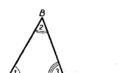
molecule at the same time, but only with its small section, which is called "antigenic determinant" or "epitope".
Distinguish linear, or sequential, antigenic determinants (for example, the primary amino acid sequence of the peptide chain) and superficial, or conformational(located on the surface of the antigen molecule and resulting from a secondary or higher conformation). In addition, there are terminal epitopes(located at the ends of the antigen molecule) and central. Also determine "deep", or hidden, antigenic determinants that appear during the destruction of the biopolymer.
The size of the antigenic determinant is small, but can vary. It is determined by the characteristics of the antigen-receptor part of the immunity factor, on the one hand, and the type of epitope, on the other. For example, the antigen-binding region of the immunoglobulin molecule (both serum and B-lymphocyte receptor) is capable of recognizing a linear antigenic determinant formed by only 5 amino acid residues. The conformational determinant is somewhat larger compared to the linear one - its formation requires 6-12 amino acid residues. The receptor apparatus of T-lymphocytes is focused on antigenic determinants that are different in structure and size. In particular, the killer T cell requires a nanopeptide included in the MHC class I to detect foreignness; When recognizing “friend or foe”, T-helper requires an oligopeptide of 12-25 amino acid residues in complex with MHC class II.
The structure and composition of the epitope are critical. Replacing at least one structural element molecules leads to the formation of a fundamentally new antigenic determinant with different properties. It should also be noted that denaturation leads to the complete or partial loss of antigenic determinants or the appearance of new ones, while the specificity of the antigen is lost.
Since the molecules of most antigens are quite large in size, many antigenic parts are determined in their structure.
terminant, which are recognized by antibodies and lymphocyte clones of different specificities. Therefore, the antigenicity of a substance depends on the presence and number of antigenic determinants in the structure of its molecule.
Foreignness is a prerequisite for the implementation of antigenicity. According to this criterion, the acquired immune system differentiates potentially dangerous objects of the biological world synthesized from a foreign genetic matrix. The concept of “foreignness” is relative, since immunocompetent cells are not able to directly analyze foreign genetic code. They perceive only indirect information, which, like in a mirror, is reflected in the molecular structure of the substance.
Normally, the immune system is immune to its own biopolymers. If a reaction occurs to any biopolymer in a macroorganism, then, accordingly, it acquires foreign features and is no longer perceived by the immune system as "mine". A similar event can occur in some pathological conditions as a result of dysregulation of the immune response (see “autoantigens”, “autoantibodies”, “autoimmunity”, “autoimmune diseases”).
Alienity is directly dependent on the “evolutionary distance” between the recipient organism and the donor of antigens. The further organisms are separated from each other in phylogenetic development, the greater the foreignness and, therefore, immunogenicity of their antigens in relation to each other. This property is used by biologists and paleontologists (when studying phylogeny, clarifying classification, etc.), forensic experts and criminologists (establishing blood relationships, evidence, falsification of food products, etc.).
Alienity is noticeably manifested even between individuals of the same species. It was noted that single amino acid substitutions, which form the basis of intraspecific polymorphism, are effectively recognized by antibodies in serological reactions.
At the same time, antigenic determinants of even genetically unrelated animals or
structurally different biopolymers may have a certain similarity. In this case, their antigens are able to specifically interact with the same immune factors. These antigens are called cross-reacting. The described phenomenon is characteristic, for example, of albumins, collagens, myoglobins of various animal species. Similarities were also found between the antigenic determinants of streptococcus, myocardial sarcolemma and the basement membrane of the kidneys, Treponema pallidum and lipid extract from the myocardium of cattle, the causative agent of plague and human erythrocytes O (I) blood group. The phenomenon when one microbe is masked by the antigens of another microbe or macroorganism for “protection” from immune factors is called antigenic mimicry.
10.1.2.2. Immunogenicity
Immunogenicity - the potential ability of an antigen to cause a specific protective reaction towards itself in the macroorganism. The degree of immunogenicity depends on a number of factors, which can be grouped into three groups:
1. Molecular features of the antigen;
2. Antigen clearance in the body;
3. Reactivity of the macroorganism.
The first group of factors includes nature, chemical composition, molecular weight, structure and some other characteristics.
Immunogenicity largely depends on nature antigen. It is known that proteins and polysaccharides have the most pronounced immunogenic properties, while nucleic acids and lipids, on the contrary, are weakly immunogenic. At the same time, their copolymers: LPS, glycoproteins, lipoproteins, are capable of sufficiently activating the immune system and therefore occupy an intermediate position in the degree of immunogenicity.
The degree of immunogenicity has a certain influence chemical composition antigen molecules. In particular, the diversity of their amino acid composition is important for the immunogenicity of proteins. It was also noted that copolymers consisting of several amino acids are more immunogenic than those consisting of a single amino acid. "Monotonic" polypeptides, post-
made from one amino acid, practically do not activate the immune system. The presence of aromatic amino acids, such as tyrosine and tryptophan, in the structure of the protein molecule significantly increases immunogenicity.
The optical isomerism of the amino slots that make up the protein molecule is also important. Peptides built from L-amino acids are easily susceptible to enzymatic degradation and are highly immunogenic. A polypeptide chain built from dextrorotatory amino acid isomers, on the contrary, is slowly cleaved by enzymes of the macroorganism and can exhibit only limited immunogenicity when administered in very small doses, since high doses of such compounds quickly lead to the development of immunological tolerance (see Chapter 11, Section 11.6 ).
Despite the apparent equivalence of antigenic determinants in terms of immunogenicity, there is a certain hierarchy in their spectrum. It is manifested by the fact that epitopes differ in their ability to induce an immune response. Therefore, when immunized with a certain antigen, the resulting spectrum of antibodies will be dominated by immunoglobulins specific to individual antigenic determinants. This phenomenon is called immunodominance. According to modern concepts, immunodominance is caused by differences in the affinity of epitopes to antigen-presenting histocompatibility complexes.
Of great importance size And molecular mass antigen. Although proteins are good at stimulating the immune system, small polypeptide molecules with a molecular weight of less than 5 kDa are generally poorly immunogenic. The minimum estimated size of an oligopeptide capable of inducing an immune response is 6-12 amino acid residues with a molecular weight of about 450 Da. As the size of the peptide increases, its immunogenicity increases. Theoretically, there is a certain relationship between these parameters, but in practice it is not always fulfilled due to the influence of extraneous factors. For example, with equal molecular weight (about 70 kDa), albumin is a stronger antigen than hemoglobin.
For polysaccharides, approximately the same dependencies remain as for peptide anti-
genes. For example, dextran, which is used in the clinic for transfusion therapy, practically does not show any immunogenicity - its molecular weight is about 75 kDa. At the same time, a polysaccharide with a molecular weight of 600 kDa induces an immune response in the human body quite well. It is noteworthy that on nucleic acids the described patterns practically do not apply.
The degree of immunogenicity is also influenced spatial structure antigen. The presence of an os-helix, branched side chains, and a high density of structurally identical epitopes in the structure of the antigen turned out to be extremely important.
It has been experimentally proven that highly dispersed colloidal solutions of antigen poorly induce an immune response. Aggregates of molecules and corpuscular antigens - whole cells (erythrocytes, bacteria, etc.) are much more immunogenic. This is due to the fact that corpuscular and highly aggregated antigens are better phagocytosed than individual molecules.
Importance spatial structure antigen is also emphasized by the fact that the fibrillar protein collagen, which has a large molecular weight (about 330 kDa), has significantly less immunogenicity compared to a globular protein such as albumin, which is almost 5 times lighter.
The steric stability of the antigen molecule also turned out to be significant. When collagen is denatured to gelatin, along with the conformational “rigidity” of the structure of the molecule, its immunogenicity almost completely disappears. Therefore, gelatin solutions are widely used for parenteral administration.
One more an important condition immunogenicity is solubility antigen. For example, such high-molecular proteins as keratin, melanin, natural silk, as well as other high-polymer compounds, cannot be obtained in the form of a colloidal solution in the normal state, and they are not immunogens. Thanks to this property, horsehair, silk, catgut and others are used in clinical practice to restore the integrity of organs and
fabrics. Therefore, the inflammatory reaction at the site of the suture or reposition should not be considered as an immunological conflict provoked by the suture material.
Second group of factors associated with the dynamics of antigen entry into the body and its elimination. Thus, the dependence of the immunogenicity of an antigen on way his introduction. This property is due to the anatomical and topographical features of the structure and development of the immune system at the sites of antigen application, as well as the biological nature of the immunogen and is necessarily taken into account when vaccination or immunization. For example, taking into account the tropism of the antigen, the vaccine against polio is administered orally, against anthrax - cutaneously, BCG - intradermally, DPT - subcutaneously, against tetanus - intramuscularly.
Affects the immune response quantity incoming antigen: the more of it, the more pronounced the immune response. However, an overdose of an antigen causes the opposite reaction - immunological tolerance. There is a logarithmic relationship between the amount of antigen and the strength of the immune response in a certain segment (interval) of doses, expressed by antigenicity equation(A. A. Vorobyov, A. V. Markovich):
lgH = alpha+ betalgD,
where al and be are coefficients characterizing, respectively, the nature of the antigen and the immunoreactivity of the macroorganism; N - strength of the immune response; D - amount of antigen.
Third group combines factors that determine the dependence of immunogenicity on the state of the macroorganism. In this regard, hereditary factors come to the fore. It is well known that the result of immunization is to a certain extent related to the genotype of the individual. There are genera and species of animals that are sensitive and insensitive to certain antigens and are used in laboratory work. For example, rabbits and rats show little or no reaction to certain bacterial antigens that can cause an extremely strong immune response in a guinea pig or mouse.
Even within a species, groups of closely related individuals can be distinguished (for example,
delirious strains of animals) that will respond differently to the administered antigen. In the course of a hybridological study, it was established that the strength of the immune response to a simple antigen in mice is determined by one gene and has a dominant mode of inheritance. The immune response to antigens of complex structure has multigene control. Moreover, in mice and guinea pigs, the association of the strength of the immune response with the genes of the major histocompatibility complex is clearly visible. In the human population, significant (tens and hundreds of times) interindividual differences in sensitivity to vaccines are also known - immunologically reactive and immunologically inert individuals are distinguished.
However, as studies have shown, along with genetic predisposition, the functional state of the macroorganism is also of no small importance - its psycho-emotional and hormonal background, the intensity of metabolic processes, etc. This determines the different levels of sensitivity to the same antigen, as in one individual at different ages. periods, and population heterogeneity as a whole.
Thus,
Immunogenicity is an important property of an antigen, which must be taken into account not only in scientific research. With immunogenicity, or rather with the individual reactivity of the macroorganism to
The introduction of an antigen causes population vaccination problems. Due to the difficulty of selecting an individual vaccine dose
I of the drug, those doses, methods and forms of its administration are used that provide the highest percentage of positive reactions in the population as a whole. It is believed that in order to prevent or stop the development of the epidemic process, it is necessary that the community has immunity. 45 % vaccinated.
The immunogenicity of an antigen can be controlled by modifying the factors listed above. There are groups of substances:
adjuvants And immunomodulators,- which are capable of nonspecifically enhancing this property of the antigen. This effect is widely used in the creation of vaccines, immunotherapy, immunoprophylaxis and research work.
10.1.2.3. Specificity
Specificity is the ability of an antigen to induce an immune response to a strictly defined epitope. This property is due to the peculiarities of the formation of the immune response - complementarity of the receptor apparatus of immunocompetent cells to a specific antigenic determinant is necessary. Therefore, the specificity of an antigen is largely determined by the properties of its constituent epitopes. However, one should take into account the arbitrary boundaries of epitopes, their structural diversity and the heterogeneity of clones with antigen-reactive lymphocyte specificity. As a result The body always responds to antigenic stimulation with a polyclonal immune response. It is estimated that up to one hundred different clones of effector lymphocytes simultaneously react to individual antigenic determinants. This results in a wide range of variations in the affinity of specific immunoglobulins, and such immunoglobulins are called polyclonal.
10.1.3. Classification of antigens Based on individual characteristic properties, the entire variety of antigens can be divided into several classification groups:
By origin,
By nature,
According to the molecular structure,
According to the degree of immunogenicity,
According to the degree of foreignness,
According to the direction of activation and availability of the immune response.
Based on their origin, antigens are distinguished between exogenous (arising outside the body) and endogenous (arising inside the body). Among endogenous ones, special attention is deserved auto- And neoantigens.
Autogenous antigens (autoantigens), or antigens of one's own body, are
structurally unchanged molecules synthesized in the body under physiological conditions. Normally, autoantigens do not cause a reaction of the immune system due to the formed immunological tolerance(immunity) or their inaccessibility to contact with immunity factors - these are the so-called behind-barrier antigens. When tolerance is broken or the integrity of biological barriers is violated (the most common cause is injury), the components of the immune system begin to specifically respond to autoantigens by producing specific immune factors (autoantibodies, a clone of autoreactive lymphocytes).
It is necessary to distinguish from autoantigens neoantigens, that arise in the body as a result of mutations. After modification, the molecules acquire foreign features.
By nature: biopolymers of protein (proteids) and non-protein nature (polysaccharides, lipids, lipopolysaccharides, nucleic acids, etc.).
According to the molecular structure: globular (the molecule has a spherical shape) and fibrillar (thread-shaped).
According to the degree of immunogenicity: complete and inferior. Full antigens have pronounced antigenicity and immunogenicity - the immune system of a sensitive organism reacts to their introduction by producing immunity factors. Such substances, as a rule, have a fairly large molecular weight (more than 10 kDa), a large molecule (particle) size in the form of a globule, and interact well with immune factors.
Defective antigens, or haptens(the term was proposed by K. Landsteiner), on the contrary, are not capable of being introduced into normal conditions induce an immune response in the body, as they have extremely low immunogenicity. However, they have not lost their antigenic property, which allows them to specifically interact with ready-made immune factors (antibodies, lymphocytes). Most often, haptens are low molecular weight compounds (molecular weight less than 10 kDa).
Under certain conditions, it is possible to force the immune system of the macroorganism
specifically respond to the hapten as a full-fledged antigen and produce immunity factors. To do this, it is necessary to artificially enlarge the hapten molecule - to connect it with a strong bond to a sufficiently large protein molecule. The carrier protein molecule is called schlepper(from German schlepper - tow). The conjugate synthesized in this way will have all the properties of a full-fledged antigen and, when introduced into the body, will cause the production of antibodies or a clone of lymphocytes specific to the hapten part of the complex. In this case, the specificity of the conjugate molecule is determined by the hapten part, and the immunogenicity is determined by the carrier protein.
Using conjugates for immunization, antibodies to hormones, drugs and other low-immunogenic compounds are obtained. Diagnostics, diagnostic kits and immunosorbents created on the basis of antibodies to low-molecular substances have significantly expanded the capabilities and increased the efficiency of laboratory diagnostics and pharmacotherapy, as well as the synthesis and isolation of highly pure bioorganic compounds.
According to the degree of foreignness: xeno-, allo- and isoantigens. Xenogeneic antigens (or heterologous) - common to organisms at different stages of evolutionary development, for example, belonging to different genera and species. For the first time, the phenomenon of commonality of a number of antigens in animals of different species was noted by D. Forsman (1911). The scientist immunized the rabbit with a suspension of guinea pig organs. It turned out that the immune serum obtained during the experiment was able to interact not only with guinea pig antigens, but also agglutinate sheep red blood cells. It was later found that the guinea pig and the sheep have a number of structurally similar antigenic determinants that cross-react. Subsequently, the list of such xenogeneic antigens was expanded by tens and hundreds of pairs and even triplets, which were formed by both warm- and cold-blooded animals, plants and microbes. All these antigens received a generalized name
rank Forsman antigens. Currently, Forsman antigens are considered from a historical perspective, and the study of heteroantigens is widely used in forensic medicine, paleontology and other areas of medicine and natural science.
Allogeneic antigens (or group) - common to genetically unrelated organisms, but belonging to the same species. Based on alloantigens, the general population of organisms can be divided into separate groups. An example of such antigens in humans are blood group antigens (ABO system, etc.) and many others. Allogeneic tissues during transplantation are immunologically incompatible - they are rejected or lysed by the recipient. Microbes can be divided into serogroups based on group antigens. It has great importance for microbiological diagnostics (for example, Kaufman-White classification of Salmonella) and epidemiological forecasting.
Isogenic antigens (or individual) - common only to genetically identical organisms, for example, identical twins, inbred lines of animals. Isografts have almost complete immunological compatibility and are not rejected by the recipient during transplantation. An example of such antigens in the human population are histocompatibility antigens, and in bacteria - typical antigens that do not give further cleavage.
Within an individual organism, in certain anatomical and morphological formations (for example, organs or tissues), antigens specific to them are found, which are no longer found in other organs and tissues. These are, for example, carcinoembryonic antigens (alpha-fetoprotein, transferrin). These antigens are collectively called organo- And tissue-specific.
A separate classification criterion is the direction of activation and the availability of the immune response in response to the introduction of the antigen. Depending on the physicochemical properties of the substance, the conditions of its introduction, the nature of the reaction and the reactivity of the macroorganism, there are immunogens, tolerogens And allergens.
Immunogens when entering the body, they are capable of inducing a productive reaction of the immune system, which ends in the production of immune factors (antibodies, antigen-reactive clones of lymphocytes). In clinical practice, immunogens are used for immunodiagnosis, immunotherapy, and immunoprophylaxis of many pathological conditions.
Tolerogen is the exact opposite of an immunogen. When interacting with the acquired immune system, it causes the inclusion of alternative mechanisms, leading to the formation of immunological tolerance or unresponsiveness to the epitopes of a given tolerogen (see section 11.6). Tolerogen, as a rule, is characterized by monomerism, low molecular weight, high epitope density and high dispersity (non-aggregation) of colloidal solutions. Tolerogens are used for the prevention and treatment of immunological conflicts and allergies by inducing artificial unresponsiveness to individual antigens.
Allergen also affects the acquired immune system. However, unlike an immunogen, the effect it produces creates a pathological reaction of the body in the form hypersensitivity immediate or delayed type (see section 11.4). In its properties, an allergen does not differ from an immunogen. In clinical practice, allergens are used to diagnose infectious and allergic diseases.
Among immunogens, two groups of antigens are distinguished, differing in the need to involve T lymphocytes in the induction of an immune response. This - T-dependent And T-independent antigens. The immune reaction in response to the introduction of a T-dependent antigen is realized with the obligatory participation of T-lymphocytes (T-helpers). Most of the known antigens are T-dependent. At the same time, the development of an immune response to T-independent antigens does not require the involvement of T helper cells. These antigens are capable of directly stimulating B lymphocytes to antibody production, differentiation and proliferation, as well as inducing an immune response in athymic individuals.
animals. T-independent antigens have a relatively simple structure. These are large molecules with a molecular weight of more than 10^3 kDa, they are polyvalent and have monotonously repeating sequences with numerous epitopes of the same type. T-independent antigens have a mitogenic effect and are capable of inducing a polyclonal reaction. Examples include the polymeric form of flagellin (the contractile protein of bacterial flagella), L PS, tuberculin, copolymers of D-amino acids, etc.
It is necessary to distinguish from T-independent antigens superantigens. This is a conventional term coined to designate a group of substances, mainly of microbial origin, that can nonspecifically cause a polyclonal reaction. In the body, bypassing the natural processing of the antigen, the whole superantigen molecule is capable of interfering with the cooperation of the antigen-representing cell and the T-helper cell and disrupting the recognition of “friend or foe”. It has been established that the superantigen molecule independently binds to the intercellular complex “class II histocompatibility antigen - T-cell receptor” and generates a false recognition signal for a foreign substance. The process of nonspecific activation simultaneously involves a huge number of T-helpers (up to 20% of the total mass or more), hyperproduction of cytokines occurs, followed by polyclonal activation of lymphocytes, their massive death due to apoptosis and the development of secondary functional immunodeficiency.
To date, superantigen properties have been found in staphylococcal enterotoxin, proteins of Epstein-Barr viruses, rabies, HIV and some other microbial substances.
10.1.4. Antigens of the human body
The study of alloantigenic properties of tissues began with K. Landsteiner, who in 1900 discovered the system of group antigens of erythrocytes (ABO). The human body produces many different antigens. As biological objects, they are needed not only for the full development and functioning of the entire organism as a whole,
but also carry important information, so necessary for clinical and laboratory diagnostics in determining the immunological compatibility of organs and tissues in transplantology, as well as in scientific research.
From the standpoint of clinical medicine, the greatest interest and importance among group-specific (allogeneic) antigens are blood group antigens, among individually specific (isogenic) antigens - histocompatibility antigens, and in the group of organo- and tissue-specific antigens - carcinoembryonic antigens.
10.1.4.1. Human blood group antigens
Human blood group antigens are easily detected on the membrane of red blood cells, which is why they are called "erythrocyte antigens". To date, more than 250 different erythrocyte antigens are known.
The antigens of the AB0 and Rh system (Rh factor) are of the most important clinical importance: they must be taken into account when carrying out blood transfusion therapy, organ and tissue transplantation, prevention and treatment of immunoconflict complications of pregnancy, etc.
Antigens of the ABO system are located on the outer membrane of all human blood cells and tissues, but are most pronounced on red blood cells. In addition, in most people (80%) these antigens are found in blood plasma, lymph, mucous secretions and other biological fluids. ABO system antigens are synthesized by nucleated red blood cell precursors and many other cells of the body. They are freely secreted into the intercellular space and therefore can appear on the cell membrane either as a product of cellular biosynthesis or as a result of sorption from intercellular fluids.
Antigens of the ABO system are highly glycosylated peptides: 85% are carbohydrate parts and 15% are polypeptide parts. The peptide component consists of 15 amino acid residues. It is constant for all ABO blood groups and is immunologically inert. The immunogenicity of an ABO system antigen molecule is determined by its carbohydrate part.
In the ABO antigen system, there are three variants of antigens that differ in the structure of the carbohydrate part: H, A and B. The basic molecule is the H antigen, the specificity of which is determined by three carbohydrate residues. Antigen A has an additional, fourth carbohydrate residue in its structure - N-acetyl-D-galactose, and antigen B - D-galactose. Antigens of the ABO system have independent allelic inheritance, which determines the presence of 4 blood groups in the population: 0(1), A ( II), B (III) and AB (IV). In addition, A and B antigens have several allotypes (for example, A1, A2 , A3... or B1, B 2, B 3...), which occur in the human population with varying frequencies.
The patient's group affiliation is determined by the ABO antigen system in an agglutination reaction - the patient's red blood cells are tested with specific group antisera. However, given the high population polymorphism of this antigenic system, before blood transfusion a biological test is required to determine the compatibility of the recipient and the donor blood product. An error in determining group affiliation and transfusion of a patient with incompatible blood group, as a rule, leads to the development of an acute condition - intravascular hemolysis, up to hemolytic shock and death of the patient.
The second most important system of erythrocyte antigens is Rh system (Rh) - so-called Rh antigens or Rh factors. These antigens are synthesized by red blood cell precursors and are found primarily on red blood cells because they are insoluble in body fluids. By chemical structure Rh antigen is a heat-labile lipoprotein. There are 6 varieties of this antigen. Genetic information about its structure is found in numerous alleles of three linked loci (D/d, C/c, E/e). Depending on the presence or absence of the Rh antigen, two groups are distinguished in the human population: Rh-positive and Rh-negative individuals.
Matching the Rh antigen is important not only for blood transfusion, but also for the course and outcome of pregnancy.
During the pregnancy of a “Rh-negative” mother, a “Rh-positive” fetus may develop "Rh-conflict". This pathological condition is associated with the production of anti-Rh antibodies, which can cause an immunological conflict: miscarriage or jaundice of the newborn (intravascular immune lysis of red blood cells).
The epitope density of the antigen on the erythrocyte membrane is low. In addition, its molecule is not convenient enough to interact with antibodies. Therefore, “Rh antigens” are determined on the membrane of erythrocytes in an indirect agglutination reaction (Coombs reaction).
10.1.4.2. Histocompatibility antigens
On the cytoplasmic membranes of almost all cells of the macroorganism are found histocompatibility antigens. Most of them relate to the system major histocompatibility complex, or MNS(abbr. from English. Main Hystocompatibility Complex).
Histocompatibility antigens play a key role in the specific recognition of “self or foe” and the induction of an acquired immune response. They determine the compatibility of organs and tissues during transplantation within the same species, genetic restriction (limitation) of the immune response and other effects.
Much credit for the study of MNS as a phenomenon of the biological world belongs to J. Dosset, P. Dougherty, P. Gorer, G. Snell, R. Zinkernagel, R. V. Petrov, who became the founders immunogenetics.
MHC was first discovered in the 60s of the 20th century. in experiments on genetically pure (inbred) lines of mice when attempting interline transplantation of tumor tissues (P. Gorer, G. Snell). In mice, this complex was named H-2 and was mapped to chromosome 17.
In humans, MHC was described somewhat later in the works of J. Dosset. He was designated as HLA(abbr. from English. Human Leukocyte Antigen), since it is associated with leukocytes. HLA biosynthesis is determined by genes
localized in several loci of the short arm of chromosome 6.
MHC has a complex structure and high polymorphism. By chemical nature, histocompatibility antigens are glycoproteins firmly associated with the cytoplasmic membrane of cells. Their individual fragments have structural homology with immunoglobulin molecules and therefore belong to a single superfamily. There are two main classes of MHC molecules. It is conventionally accepted that class I MHC induces predominantly a cellular immune response, and class II MHC induces a humoral response. The main classes combine many structurally similar antigens, which are encoded by many allelic genes. In this case, no more than two types of products of each MHC gene can be expressed on the cells of an individual, which is important for maintaining population heterogeneity and the survival of both an individual and the entire population as a whole.
MHC class I consists of two non-covalently linked polypeptide chains with different molecular weights: a heavy alpha chain and a light beta chain (Fig. 10.1). The alpha chain has an extracellular region with a domain structure (alfa1-, a2- and a3-domains), transmembrane and cytoplasmic. The beta chain is a beta-2 microglobulin that sticks to the a3 domain after expression of the alpha chain on the cytoplasmic membrane of the cell.
The alpha chain has a high sorption capacity for peptides. This property is determined by the all- and a2-domains, which form the so-called “Bjorkman gap” - a hypervariable region responsible for the sorption and presentation of antigen molecules. The “Björkman gap” of MHC class I contains a nanopeptide, which in this form is easily detected by specific antibodies.
The process of formation of the MHC class I-antigen complex occurs intracellularly continuously. It includes any endogenously synthesized peptides, including viral ones. The complex is initially assembled in the endoplasmic reticulum, where, with the help of a special protein, proteasomes, peptides are transferred from the cytoplasm. The peptide included in the complex imparts structural stability to MHC class I. In its absence, the function of a stabilizer is performed chaperone (calnexin).
MHC class I is characterized by a high rate of biosynthesis - the process is completed in 6 hours. This complex is expressed on the surface of almost all cells, except for erythrocytes (there is no biosynthesis in anucleate cells) and villous trophoblast cells (“prevention” of fetal rejection). The density of MHC class I reaches 7000 molecules per cell, and they cover about 1 % its surface. The expression of the molecules is markedly enhanced by cytokines, such as gama-interferon.
Currently, there are more than 200 different HLA class I variants in humans. They are encoded by genes mapped to three main subloci of chromosome 6 and are inherited and expressed independently: HLA-A, HLA-B and HLA-C. Locus A unites more than 60 variants, B - 130, and C - about 40.
Typing an individual according to HLA class I is carried out on lymphocytes using serological methods - in a microlymphocytolysis reaction with specific sera. For diagnosis, polyclonal specific antibodies are used, found in the blood serum of multiparous women, patients who received massive blood transfusion therapy, as well as monoclonal ones.
Taking into account the independent inheritance of sublocus genes, an infinite number of non-repeating combinations of HLA class I are formed in the population. Therefore, each person is strictly unique in terms of the set of histos-carrying antigens, with the only exception being identical twins, who are absolutely similar in their set of genes. The main biological role of HLA class I is that they determine biological individuality (“biological passport”) and are “self” markers for immunocompetent cells. Infection of a cell with a virus or mutation changes the structure of HLA class I. The MHC class I molecule containing foreign or modified peptides has a structure atypical for a given organism and is a signal for the activation of T-killer cells (CD8 + lymphocytes). Cells that differ in class I are destroyed as foreign.
There are a number of fundamental differences in the structure and function of class II MHC. Firstly, they have more complex structure. The complex is formed by two non-covalently linked polypeptide chains (alpha chain and beta chain) having a similar domain structure (Fig. 10.1). The alpha chain has one globular region, and the beta chain has two. Both chains, as transmembrane peptides, consist of three sections - extracellular, transmembrane and cytoplasmic.
Secondly, the “Björkman gap” in class II MHC is formed simultaneously by both chains. It accommodates a larger oligopeptide (12-25 amino acid residues), and the latter is completely “hidden” inside this gap and in this state is not detected by specific antibodies.
Third, MHC class II includes a peptide taken up from the extracellular environment by endocytosis, and not synthesized by the cell itself.
Fourth, MHC class II is expressed on the surface of a limited number of cells: dendritic, B-lymphocytes, T-helper cells, activated macrophages, mast, epithelial and endothelial cells. The detection of MHC class II on atypical cells is currently regarded as immunopathology.
Biosynthesis of MHC class II occurs in the endoplasmic reticulum, the resulting dimeric complex is then integrated into the cytoplasmic membrane. Before the peptide is included in it, the complex is stabilized by a chaperone (calnexin). MHC class II is expressed on the cell membrane within an hour after endocytosis of the antigen. Expression of the complex can be enhanced by interferon-ga and reduced by prostaglandin E2.
In mice, the histocompatibility antigen is called la-antigen, and in humans, by analogy, it is called class HLAII.
According to available data, the human body is characterized by extremely high polymorphism of HLA class II, which is largely determined by the structural features of the beta chain. The complex includes products of three main loci: HLA DR, DQ and DP. At the same time, the DR locus unites about 300 allelic forms, DQ - about 400, and DP - about 500.
The presence and type of class II histocompatibility antigens are determined by serological (microlymphocytotoxic test) and cellular reactions immunity (mixed culture of lymphocytes, or MCL). Serological typing of MHC class II is carried out on B-lymphocytes using specific antibodies found in the blood serum of multiparous women, patients who received massive blood transfusion therapy, as well as those synthesized by methods genetic engineering. Testing in the SCL makes it possible to identify minor components of MHC class II that are not detectable serologically. Recently, PCR has been increasingly used.
Biological role MHC class II is extremely large. In fact, this complex is involved in the induction of the acquired immune response. Fragments of the antigen molecule are expressed on the cytoplasmic membrane of a special group of cells, which is called antigen presenting cells (APCs). This is an even narrower circle among cells capable of synthesizing MHC class II. The dendritic cell is considered the most active APC, followed by the B lymphocyte and macrophage. The structure of class II MNS with incl.
The peptide contained in it, in combination with co-factor molecules of CD antigens, is perceived and analyzed by T helper cells (CD4 + lymphocytes). If a decision is made about the foreignness of a peptide included in the MHC class II, the T-helper begins the synthesis of the corresponding immunocytokines, and the mechanism of a specific immune response is activated. As a result, proliferation and final differentiation of antigen-specific lymphocyte clones and the formation of immune memory are activated.
In addition to the histocompatibility antigens described above, class III MHC molecules have been identified. The locus containing the genes encoding them is wedged between class I and class II and separates them. MHC class III includes some complement components (C2, C4), heat shock proteins, tumor necrosis factors, etc.
10.1.4.3. Tumor-associated antigens
The first indications of the presence of specific antigens in tumors date back to the 40s of the 20th century. In 1948-1949 L.A. Zilber, a prominent Russian microbiologist and immunologist, when developing the viral theory of cancer, proved the existence of an antigen specific to tumor tissue. Later, in the 60s of the 20th century, G. I. Abelev (in experiments on mice) and Yu. S. Tatarinov (while examining people) discovered an embryonic version of serum albumin in the blood serum of patients with primary liver cancer - alpha fetoprotein. To date, tumor-associated antigens have been discovered and characterized for many tumors, and their genes have even been cloned. However, not all tumors contain specific marker antigens, and not all markers have strict tissue specificity.
Tumor-associated antigens are classified by location and genesis. By location they distinguish whey, secreted by tumor cells into the intercellular environment, and membrane The latter were called tumor-specific transplantation antigens, or TSTA (abbreviation for English. Tumor-specific transplantation antigen).
Depending on the nature they distinguish viral, embryonic, normal overexpressed And mutant antigens associated with tumors. Viral Tumor-associated antigens are essentially proteins of oncoviruses. Embryonic antigens are normally synthesized in the embryonic period. This is, for example, alpha-fetoprotein (see above); normal testicular protein, MAGE 1, 2, 3, etc. - markers of normal testes, as well as melanoma, breast cancer, etc.; human chorionic gonadotropin is normally synthesized in the placenta, as well as in choriocarcinoma and other tumors. In melanoma large quantities the normal enzyme tyrosinase is synthesized.
From mutant proteins, it is worth noting the Ras protein, characteristic of many tumors, a GTP-binding protein involved in transmembrane signal transmission. Markers of breast and pancreatic cancer, intestinal carcinoma are modified mucins (MUC 1, 2, etc.).
From general properties tumor-associated antigens, it should be noted that most of them are products of the expression of genes that are normally turned on only in the embryonic period. They are weak immunogens, although in some cases can induce a reaction of cytotoxic T-lymphocytes (T-killers) and are recognized as part of MHC (HLA) class I molecules. Specific antibodies directed against tumor-associated antigens, in essence, do not inhibit tumor growth, but, on the contrary, cause immunosuppression.
10.1.4.4. CD antigens
Group antigens are found on the cell membrane, uniting cells that have similar morphofunctional characteristics or are at a certain stage of development. These marker molecules are called cell differentiation cluster antigens, or CD antigens (abbreviated from the English. Cell Differentiation Antigens, or Cluster Definition). Structurally, they are glycoproteins, many of which belong to the immunoglobulin superfamily.
CD antigens are used to identify differences in groups of cells, of which the most widely used markers are immunocompetent cell markers. For example, CD3 is expressed on the population of T-lymphocytes, CD4 is characteristic of the subpopulation of T-helper cells, and CD8 is characteristic of cytotoxic T-lymphocytes of killer T-cells. CDlla is found on the cytoplasmic membranes of mono- and granulonites, and CDllb is found on natural killer cells. CD19-22 are markers of B lymphocytes.
The list of CD markers is quite extensive, it has about 200 options. The main CD markers of cells involved in the immune response are presented in Table. 10.1. Information about the structure is encoded in various parts of the genome, and expression depends on the stage of cell differentiation and its functional state.
CD antigens have diagnostic value in the clinic of immunodeficiency conditions, as well as in research work. Typing of CD markers is carried out in serological reactions using monoclonal antibodies (immunofluorescence reaction, cytotoxic test, etc.).
10.1.5. Antigens of microbes
Several types of antigens are determined in the structure of microbes. Moreover, the antigenic composition of a microbe largely depends on its evolutionary and taxonomic position. The antigens of bacteria, viruses, fungi and protozoa have fundamental differences.
However, microbial antigens may be common to certain systematic categories. Thus, there are antigens characteristic of entire families, genera and species. Within species, serological groups (serogroups), variants (serovars) or types (serotypes) can be distinguished. Microbial antigens are used to obtain vaccines and serums necessary for the diagnosis, prevention and treatment of infectious or allergic diseases, as well as in diagnostic reactions.
10.1.5.1. Antigens of bacteria
In the structure of a bacterial cell, flagellar, somatic, capsule and some other antigens are distinguished (Fig. 10.2). Flagellates, or H-antigens are localized in the locomotor apparatus of bacteria - their flagella. They are epitopes of the contractile protein flagellin. When heated, flagellin denatures and H-antigen loses its specificity. Phenol has no effect on this antigen.
Somatic, or O-antigen, associated with the bacterial cell wall. It is based on LPS. O-antigen exhibits thermostable properties - it is not destroyed by prolonged boiling. However, the somatic antigen is susceptible to the action of aldehydes (for example, formaldehyde) and alcohols, which disrupt its structure.
If you immunize an animal with live bacteria that have flagella, antibodies will be produced that are directed simultaneously against O- and H-antigens. Introducing a boiled culture to an animal stimulates the biosynthesis of antibodies to a somatic antigen. Bacterial culture treated with pheno-
scrap will cause the formation of antibodies to flagellar antigens.
Capsule, or K-antigens located on the surface of the cell wall. Found in capsule-forming bacteria. As a rule, K-antigens consist of acidic polysaccharides (uronic acids). At the same time, in the anthrax bacillus, this antigen is built from polypeptide chains. Based on their sensitivity to heat, there are three types of K-antigen: A, B, and L. The greatest thermal stability is characteristic of type A; it does not denature even with prolonged boiling. Type B can withstand short heating (about 1 hour) up to 60 °C. Type L degrades quickly at this temperature. Therefore, partial removal of the K-antigen is possible by prolonged boiling of the bacterial culture.
On the surface of the causative agent of typhoid fever and other enterobacteria that are highly virulent, a special version of the capsular antigen can be found. It got the name virulence antigen, or Vi-antigen. Detection of this antigen or antibodies specific to it is of great diagnostic importance.
Bacterial bacteria also have antigenic properties. protein toxins, enzymes and some other proteins that are secreted by bacteria in environment(for example, tuberculin). When interacting with specific antibodies, toxins, enzymes and other biologically active molecules of bacterial origin lose their activity. Tetanus, diphtheria and botulinum toxins are among the strong full-fledged antigens, so they are used to obtain toxoids for human vaccination.
The antigenic composition of some bacteria contains a group of antigens with highly expressed immunogenicity, whose biological activity plays a key role in the formation of the pathogenicity of the pathogen. The binding of such antigens by specific antibodies almost completely inactivates the virulent properties of the microorganism and provides immunity to it. The described antigens are called protective. For the first time, a protective antigen was discovered in purulent
nom discharge of the carbuncle caused by the anthrax bacillus. This substance is a subunit of a protein toxin, which is responsible for the activation of other, actually virulent subunits - the so-called edematous And lethal factors.
10.1.5.2. Antigens of viruses
In the structure of the viral particle, several groups of antigens are distinguished: nuclear(or cow) capsid(or shell) and supercapsid. On the surface of some viral particles there are special V-antigens- hemagglutinin and the enzyme neuraminidase. Viral antigens differ in origin. Some of them are virus-specific. Information about their structure is mapped in the nucleic acid of the virus. Other viral antigens are components of the host cell (carbohydrates, lipids). they are captured in the outer shell of the virus at its birth by budding.
The antigenic composition of the virion depends on the structure of the viral particle itself. The antigenic specificity of simply organized viruses is associated with ribo- and deoxyribonucleoproteins. These substances are highly soluble in water and are therefore designated as S-antigens (from Lat. solutio- solution). In complex viruses, part of the antigen is associated with the nucleocapsid, and the other is localized in the outer shell - the supercapsid.
The antigens of many viruses are highly variable. This is due to the constant mutation process that the genetic apparatus of the viral particle undergoes. Examples include influenza virus and human immunodeficiency viruses.
10.1.6. Processes occurring with the antigen in the macroorganism
The process of antigen penetration and its contact with the immune system occurs in stages and has its own dynamics over time. Moreover, at each stage of the appearance and spread in the macroorganism, the antigen faces powerful resistance from a developed network of various immune factors (see Table 9.3.).
There are various ways of penetration and spread of antigen in the macro-
roorganism. They can appear within the macroorganism itself (endogenous origin) or come from outside (exogenous origin). Exogenous origin suggests that the antigen can penetrate the macroorganism:
1) through defects in the skin and mucous membranes (as a result of wounds, microtraumas, insect bites, scratching, etc.);
2) by absorption in the gastrointestinal tract (endocytosis by epithelial cells);
In the body, the antigen is carried by lymph (lymphogenous pathway) and blood (hematogenous pathway) to various organs and tissues. Moreover, it is not distributed chaotically - the antigen is most often filtered in the lymph nodes, as well as in the lymphoid tissue of the liver, spleen, lungs and other organs, where it comes into contact with various immune defense factors.
The response of these factors is the inactivation and removal (elimination) of the antigen from the macroorganism. The factors of innate immunity come into play first, since this system, despite its diversity and complexity of its individual components, does not require a long time to activate. If the antigen has not been inactivated or eliminated within 4 hours, the system of acquired immune factors is activated. The effectiveness of their action is ensured by specific recognition "friend or foe" and the production of appropriate regulatory factors and immune defense (specific antibodies, clones of antigen-reactive lymphocytes).
The cumulative effect of all links and levels of the immune defense of the macroorganism, regardless of the degree of their involvement in the process, is aimed at:
1) binding and blocking biologically active sites of the antigen molecule;
2) destruction or rejection of the antigen;
3) complete utilization, isolation (encapsulation) or removal of antigen remnants from the macroorganism.
As a result, complete or partial restoration of homeostasis is achieved. At the same time, immune memory, tolerance or allergy is formed.
Lecture outline:
1. Antigens: definition, structure, basic properties.
2. Antigens of microorganisms.
3. Antigens of humans and animals.
4. Antibodies: definition, main functions, structure.
5. Classes of immunoglobulins, their characteristics.
6. Dynamics of antibody formation.
Antigens (from Greek. anti- against, genos- create; term proposed in 1899 Deutsch) - substances of various origins that carry signs of genetic foreignness and, when introduced into the body, cause the development of specific immunological reactions.
Main functions of antigens:
Induce an immunological response (synthesis of antibodies and launch of cellular immunity reactions).
They specifically interact with the resulting antibodies (in vivo and in vitro).
Provide immunological memory- the body’s ability to respond to repeated introduction of an antigen with an immunological reaction characterized by greater strength and faster development.
Determine development immunological tolerance- lack of an immune response to a specific antigen while maintaining the ability to have an immune response to other antigens.
Structure of antigens:
Antigens consist of 2 parts:
1. High molecular weight carrier (schlepper)- a high-polymer protein that determines the antigenicity and immunogenicity of the antigen.
2. Determinant groups (epitopes)- surface structures of the antigen, complementary active center antibodies or T-lymphocyte receptor and determining the specificity of the antigen. One carrier may have several different epitopes, consisting of peptides or lipopolysaccharides and located in different parts of the antigen molecule. Their diversity is achieved due to a mosaic of amino acid or lipopolysaccharide residues located on the surface of the protein.
The number of determinant groups or epitopes determines antigen valency.
Antigen valence- the number of identical epitopes on an antigen molecule, equal to the number of antibody molecules that can attach to it.
Main properties of antigens:
1. Immunogenicity- the ability to induce immunity, immunity to infection (used to characterize infectious agents).
2. Antigenicity- the ability to induce the formation of specific antibodies (a particular variant of immunogenicity).
3. Specificity- a property by which antigens differ from each other and determines the ability to selectively react with specific antibodies or sensitized lymphocytes.
Immunogenicity, antigenicity and specificity depend on many factors.
Factors determining antigenicity:
- Foreignness (heterogeneity)- the genetically determined property of the antigens of some animal species to differ from the antigens of other animal species (the farther the animals are from each other phenotypically, the more antigenicity they have in relation to each other).
- Molecular weight must be at least 10,000 daltons; with increasing molecular weight, antigenicity increases.
- Chemical nature and chemical homogeneity: the most antigenic are proteins, their complexes with lipids (lipoproteins), with carbohydrates (glycoproteins), with nucleic acids (nucleoproteins), as well as complex polysaccharides (with a mass of more than 100,000 D), lipopolysaccharides; Nucleic acids and lipids themselves are non-immunogenic due to insufficient structural rigidity.
- Structure rigidity(in addition to a certain chemical nature, antigens must have a certain rigidity of structure, for example, denatured proteins do not have antigenicity).
- Solubility(insoluble proteins cannot be in the colloidal phase and do not cause the development of immune reactions).
Factors determining immunogenicity:
Properties of antigens.
Method of antigen administration (oral, intradermal, intramuscular).
Antigen dose.
Interval between administrations.
The state of an immunized macroorganism.
The rate of destruction of the antigen in the body and its removal from the body.
Immunogenicity and antigenicity may not be the same! For example, the dysentery bacillus is highly antigenic, but no pronounced immunity against dysentery is developed.
Factors that determine specificity:
Chemical nature of the antigenic determinant.
The structure of the antigenic determinant (type and sequence of amino acids in the primary polypeptide chain).
Spatial configuration of antigenic determinants.
Types of antigens by structure:
1. Haptens (defective antigens)- this is a pure determinant group (they have a small molecular weight, are not recognized by immunocompetent cells, have only specificity, i.e. they are not able to cause the formation of antibodies, but enter into a specific reaction with them):
- simple- interact with antibodies in the body, but are not able to react with them in vitro;
- complex- interact with antibodies in vivo and in vitro.
2. Complete (conjugated) antigens- are formed when a hapten binds to a high molecular weight carrier that is immunogenic.
3. Half-haptens- these are inorganic radicals (J -, Cr -, Br -, N +) bound by protein molecules.
4. Proantigens- haptens that can attach to body proteins and sensitize them as autoantigens.
5. Tolerogens- antigens that can suppress immunological reactions with the development of a specific inability to respond to them.
Types of antigens according to the degree of foreignness:
1. Species antigens- antigens of a certain type of organism.
2. Group antigens (alloantigens)- antigens that cause intraspecific differences in individuals of the same species, dividing them into groups (serogroups in microorganisms, blood groups in humans).
3. Individual antigens (isoantigens)- antigens of a specific individual.
4. Heterogeneous (cross-reacting, xenoantigens) antigens- antigens common to organisms different types, far apart from each other:
- antigenic mimicry- long-term absence of an immunological reaction to antigens due to similarity with host antigens (microorganisms are not recognized as foreign);
- cross reactions- antibodies formed against the antigens of microorganisms come into contact with the host antigens and can cause an immunological process (for example: hemolytic streptococcus has cross-reacting antigens with the antigens of the myocardium and renal glomeruli; the measles virus has cross-reacting antigens to the myelin protein, therefore the immune reaction contributes to the demyelination of nerve fibers and the development multiple sclerosis).
Antigens of microorganisms depending on systematic position:
1. Species-specific- antigens of one type of microorganism.
2. Group-specific- antigens of one group within a species (divide microorganisms into serogroups).
3. Type-specific- antigens of one type (variant) within a species (divide microorganisms into serovars/serotypes).
Specificity - this is the ability of an antigen to interact with strictly defined antibodies or antigen receptors of lymphocytes.
In this case, the interaction does not occur with the entire surface of the antigen, but only with its small section, which is called the “antigenic determinant” or “epitope”. One antigen molecule can have from several units to several hundred epitopes of varying specificity. The number of epitopes determines the valency of the antigen. For example: egg albumin (M 42,000) has 5 epitopes, i.e. 5-valentene, thyroglobulin protein (M 680,000) - 40-valentene.
In protein molecules, the epitope (antigenic determinant) is formed by a set of amino acid residues. The size of the antigenic determinant of proteins can include from 5 - 7 to 20 amino acid residues. Epitopes that are recognized by antigen receptors of B and T lymphocytes have their own characteristics.
B-cell epitopes of conformational type (formed by amino acid residues from various parts protein molecule, but close in the spatial configuration of the protein globule) are located on the outer surface of the antigen, forming loops and protrusions. Typically, the number of amino acids or sugars in an epitope is from 6 to 8. Antigen recognition receptors of B cells recognize the native conformation of the epitope, rather than a linear sequence of amino acid residues.
T cell epitopes are a linear sequence of amino acid residues that form part of an antigen and include larger number amino acid residues compared to B-cell ones. Their recognition does not require saving the spatial configuration.
Immunogenicity - the ability of an antigen to induce immune defense of the macroorganism. The degree of immunogenicity is determined by the following factors:- Foreignness . In order for a substance to act as an immunogen, it must be recognized as “not its own.” The more foreign the antigen is, that is, the less similar it is to the body’s own structures, the stronger the immune response it causes. For example, the synthesis of antibodies to bovine serum albumin is easier to induce in a rabbit than in a goat. Rabbits belong to the order of lagomorphs and are further away in phylogenetic development from the goat and bull, which belong to the artiodactyls.
- Nature of the antigen . The most powerful immunogens are proteins. Pure polysaccharides, nucleic acids and lipids have weak immunogenic properties. At the same time, lipopolysaccharides, glycoproteins, and lipoproteins are capable of sufficiently activating the immune system.
- Molecular mass . All other things being equal, the larger molecular weight of the antigen provides greater immunogenicity. Antigens are considered good immunogens if their molecular weight is more than 10 kDa. The higher the molecular weight, the more binding sites (epitopes), which leads to an increase in the intensity of the immune response.
- Solubility. Corpuscular antigens associated with cells (erythrocytes, bacteria) are usually more immunogenic. Soluble antigens (serum albumin) may also be highly immunogenic, but are cleared more quickly. To increase the time they remain in the body, necessary for the development of an effective immune response, adjuvants (depositing substances) are used. Adjuvants are substances that are used to enhance the immune response, for example, liquid paraffin, lanolin, aluminum hydroxide and phosphate, potassium alum, calcium chloride, etc.
- Chemical structure of antigen . Increasing the number of aromatic amino acids in synthetic polypeptides increases their immunogenicity. With equal molecular weight (about 70,000), albumin is a stronger antigen than hemoglobin. At the same time, the collagen protein, whose molecular weight is 5 times greater than that of albumin and amounts to 330,000, has significantly less immunogenicity compared to albumin, which is undoubtedly due to the structural features of these proteins.
Immunogenicity (Immuno- + Greek -genēs generating, producing)
1. Small medical encyclopedia. - M.: Medical encyclopedia. 1991-96 2. First health care. - M.: Great Russian Encyclopedia. 1994 3. Encyclopedic Dictionary medical terms. - M.: Soviet encyclopedia. - 1982-1984.
See what “Immunogenicity” is in other dictionaries:
Immunogenicity is the ability of an antigen to provoke an immune response, regardless of its immune specificity. The degree of immunogenicity depends not only on the properties of the antigen molecule, but also on the conditions of introduction into the body, as well as additional ... ... Wikipedia
immunogenicity- The ability of the drug to cause an immune response. [English-Russian glossary of basic terms in vaccinology and immunization. World Health Organization, 2009] Topics vaccinology, immunization EN immunogenicityimmunogenic activity ... Directory technical translator
- (immuno + Greek genes generating, producing) the ability of a substance to cause a specific immune response with the development of immunity ... Large medical dictionary
immunogenicity- immunogenicity, and... Russian spelling dictionary
Immunogenicity- - the ability of substances to cause a specific immune response with the development of immunity ... Glossary of terms on the physiology of farm animals
- (from the Greek ἅπτω to attach) low-molecular substances that do not have immunogenicity and acquire them with an increase in molecular weight (for example, due to attachment to a special protein carrier, the so-called “schlepper”). In... ... Wikipedia
Main article: Hepatitis B Vaccine against the hepatitis B virus is an immunobiological preparation, a group of vaccines against hepatitis B, from different manufacturers. Although vaccination is only one of several ways to prevent diseases,... ... Wikipedia
- (Greek anti against + gennao create, produce) bioorganic substances that have signs of genetic foreignness (antigenicity) and when introduced into the body cause the development of an immune response. Antigenicity is not limited to proteins... Medical encyclopedia
- (lat. bovine vaccinus) preparations obtained from microorganisms or their metabolic products; are used for active immunization of people and animals for preventive and therapeutic purposes. Vaccines consist of the active principle of a specific... Medical encyclopedia
Main article: Influenza A vaccine for the prevention of influenza, a drug from the group of biological drugs that provides short-term immunity to the influenza virus, is considered one of the most effective means of prevention... ... Wikipedia
Cervarix is a recombinant adsorbed vaccine for the prevention of diseases caused by human papillomaviruses (HPV), containing the adjuvant AS04. It is a mixture of virus-like particles of recombinant surface proteins of HPV... ... Wikipedia
All vaccines, except genetically engineered ones, are heterogeneous in their antigenic composition. When corpuscular vaccines (live or killed) are administered, their decay products appear, differing in physical and chemical properties. Oligomers, monomers and low molecular weight fragments are formed. The latter are able to interact with specific receptors of immunocomponent cells without causing an immune response. In addition, very large antigen molecules with high degree valences can also be tolerogenic. Less heterogeneous are toxoids and highly purified microbial fractions used as vaccines.
The immunogenicity of full antigens included in vaccines depends on the size and polymerity of their molecules, the immunogenicity of haptens depends on their epitope density on the carrier molecule. A low-polymer antigen can cause not only a weak, but also a qualitatively different type of immune response compared to a high-polymer antigen.
From the point of view of molecular and cellular immunology, the vaccine must meet the following requirements:
- The vaccine must activate supporting cells (macrophages, dendritic cells, Langerhans cells) involved in antigen processing and presentation.
- It must contain epitopes for T and B cells, ensuring the necessary balance of humoral and cellular immunity.
- It must be easily processed, and its epitopes must be able to interact with histocompatibility antigens of class 1 and/or class II.
- It should induce the formation of regulatory cells (T-helpers), effector cells (killer cells, T-effectors of HRT, antibody-forming cells) and immunological memory cells.
An ideal vaccine must meet two basic requirements: it must be safe and highly effective. It should be administered once and provide lifelong immunity to 100% of those vaccinated. There are no such vaccines yet. Despite great strides in improving existing vaccines and developing new drugs, the duration of immunity that occurs after administration of most vaccines is short, even if the same vaccine is administered multiple times. For some vaccines it is only 1 year (Table 25). The data indicated in the table were obtained by different authors in different time and are quite conditional. It should be noted that in immunized individuals, a certain degree of specific protection remains even after the disappearance of circulating antibodies.
Table 25. Duration of immunity (based on protective antibody titers) after primary immunization
|
The strength of the immune response depends on two main factors: the properties of the macroorganism and the characteristics of the antigens used for immunization. The immunogenicity of antigens obtained from pathogens of infectious diseases is not the same. The most immunogenic are exotoxins and surface antigens of microorganisms. The immunogenicity of a vaccine largely depends on how well the antigens are chosen for the design of the drug. If its immunogenicity is insufficient, nonspecific immunostimulants (adjuvants) are used. In the practice of vaccination, aluminum hydroxide, aluminum phosphate, calcium phosphate, polyoxidonium and protein carriers are used as immunostimulants.
Difficulties in creating highly effective vaccines are also associated with the characteristics of the macroorganism, its genotype, phenotype, and the existence of two types of immunity (humoral and cellular), which are regulated by different subpopulations of helper cells (Th1 and Th2). Post-vaccination immunity consists of two types of immune reactions: humoral and cellular. The absence of circulating antibodies is not yet evidence of weak immunity, but new meeting with an antigen, the immune response develops due to immunological memory. In addition, resistance to certain types of infections is based on cellular mechanisms, so vaccines used to prevent these infections must build cellular immunity.
The immunogenicity of vaccines is the basis of their effectiveness. As a rule, the corpuscularity of vaccines (live, killed) provides the necessary immunogenicity; in other cases, it is often necessary to use additional methods to increase the immunogenicity of vaccines.
Ways to increase the immunogenicity of vaccines
- Use of optimal antigen concentration.
- Purification of vaccines from low molecular weight substances that can cause specific or nonspecific suppression of the immune response.
- Aggregation of antigen by covalent binding and other complexation methods.
- Inclusion of the maximum number of antigen epitopes in the vaccine.
- Sorption on substances that create an antigen depot (aluminum hydroxide, calcium phosphate, etc.).
- Use of liposomes (water-oil emulsion).
- Addition of microbial, plant, synthetic and other types of adjuvants.
- Binding of a weak antigen to a protein carrier (tetanus, diphtheria toxoid, etc.).
- Incorporation of antigen into microcapsules allowing release of antigen through specified interval time.
10. Improving the conditions for antigen processing and presentation. The use of histocompatibility antigens of classes 1 and 2 or antibodies to these antigens.
Approaches to creating vaccines that ensure the formation of cellular and humoral immunity are different. This is due to the participation of two regulatory cells in the immune response: Th1 and Th2. There is a certain degree of antagonism between them, although they are formed from the same type of progenitor cells. It is quite difficult to obtain a vaccine that would induce cellular immunity. In many cases, it is not possible to switch the Th2 immune response to the vaccine, which stimulates antibody production, to a Th1 cellular response.
It is critical that vaccines induce a T-dependent immune response. Otherwise, the response will be short-lived and repeated administration of the vaccine will not produce a secondary response. The primary and secondary immune response differ from each other in the dynamics of immunity formation (Fig. 11). The secondary immune response is not sufficiently expressed if a weak antigen is used for immunization, if passively introduced or actively acquired antibodies are present in the body, if the antigen is administered to a patient with immunodeficiency.
The secondary immune response is characterized by the following features:
- Earlier (compared to the primary response) development of immune reactions.
- Reducing the dose of antigen required to achieve an optimal response.
- Increased strength and duration of the immune response.
- Strengthening humoral immunity:
– increase in the number of antibody-forming cells and circulating antibodies;
– activation of Th2 and increased production of their cytokines (IL-3, 4, 5, 6, 9, 10, 13, GM-CSF, etc.);
– reduction in the period of formation of IgM antibodies, predominance of 1nSt and IgA antibodies;
– increasing the affinity of antibodies.
5. Strengthening cellular immunity:
– increase in the number of antigen-specific T-killers and T-effectors of HRT;
– activation of Th1 and increased production of their cytokines (IF-γ, FIO, IL-2, GM-CSF, etc.);
– increasing the affinity of antigen-specific T-cell receptors.
6.Increasing resistance to infection.
The body acquires the ability to quickly respond to repeated contact with an antigen thanks to immunological memory. It is characteristic of cellular and humoral immunity and depends on the formation of T- and B-memory cells. Immunological memory develops after an infection or vaccination and persists for a long time.
In some infections, antibodies remain in the serum for decades. However, the half-life of the most stable immunoglobulin is on average 25 days. Thus, the body constantly re-synthesizes specific immunoglobulin.
The duration of post-infectious immunity depends on the properties of the pathogen, the infectious dose, the state of the immune system, genotype, age and other factors. Immunity can be short-term, for example with influenza, dysentery, relapsing fever, quite long-lasting, for example with anthrax, rickettsiosis, leptospirosis, and even lifelong, for example with polio, measles, whooping cough.
Acquired immunity is a good defense against infection by the same pathogen. If the main mechanism of immunity during a given infection is the neutralization effect, then the presence of a certain level of circulating antibodies is sufficient to prevent reinfection.
To achieve lasting immunity, vaccines must be administered 2 times or more. Primary vaccination may consist of several doses of the vaccine, the intervals between doses are strictly regulated. The schedule for revaccination is more flexible; revaccination can be carried out after a year or even after several years.
The interval between vaccine administrations should be at least 4 weeks. Otherwise, less stable immunity develops. Conversely, slightly increasing the 4-week interval may enhance the secondary immune response. The maximum increase in antibody concentration during the secondary response to vaccines occurs at low initial antibody titers. A high previous level of antibodies prevents additional production of antibodies and their long-term preservation, and in some cases a decrease in antibody titers is observed.