Syndromes of lesions of the subcortical region
The defeat of the corpus callosum is characterized by mental disorders, increasing dementia, memory loss, orientation in space is disturbed, and apraxia of the left hand develops.
The thalamic Dejerine-Roussy syndrome is characterized on the opposite side by hemianesthesia, sensitive hemiataxy, and thalamic pain. There is a thalamic hand, choreo-athetoid hyperkinesis, and violent laughter and crying.
Hypothalamic syndrome consists of disorders of carbohydrate, fat, protein metabolism, disorders of the cardiovascular, respiratory and gastrointestinal systems. There may be obesity, cachexia, impotence, menstrual irregularities. Sleep and wakefulness disturbance.
With the defeat of the epithelium: accelerated puberty, increased growth, ataxia are observed.
Foreign lesion syndrome (metathalamus): damage to the external and internal geniculate bodies is characterized by hearing loss, homonymous (central and peripheral) hemianopia.
Syndromes of damage to the internal capsule: hemianesthesia, hemiplegia and hemianopsia on the opposite side. Syndrome of damage to the radiant crown: hemiparesis, hemihypesthesia, monoparesis, monoplegia with uneven damage to the arms and legs.
Parkinson's syndrome: akinesia, hypokinesia, oligokinesia, plastic hypertension of the muscles, "cog wheel", "wax doll" symptom, throwing to the sides when walking, parkinsonian marking time, slowness of thinking, paradoxical movements.
There may be an increase in postural reflexes, a quiet monotonous voice, a violation of posture and gait (the head and torso are tilted forward, the arms are bent at the elbow and wrist joints, the legs are at the knees and are slightly adducted), pallidar tremor is characteristic.
Syndrome of lesions of the striatum (hypotonic-hyperkinetic syndrome): hypotension, chorea, athetosis, choreoathetosis, facial hemispasm, facial paraspasm, hemitremor, torsion spasm, myoclonus; tics, blepharospasm, platysma spasm, torticollis. When the subthalamic nucleus (Lewis body) is damaged, hemiballismus is observed.
subcortical area
Subcortical formations are an accumulation of gray matter closest to the cerebral cortex. Caudate nucleus formed from the anterior bladder and is closer in origin to the cerebral cortex. Lenticular nucleus subdivided into shell and pale ball. Close in structure, the shell and the caudate nucleus, as well as later formations, made up the nucleus, called the striatum (striatum). The pale ball (pallidum) is an older formation, an antagonist of the striatum. The striatum and globus pallidus form a strio-pallidar system. Almond nucleus closely related to the limbic region. The meaning of the fence is unclear.
The structure of the subcortical nodes is quite complicated. Thus, the striatum is characterized by the presence of both large and small polygonal cells, characterized by chromatophilic cytoplasm and a large number of dendrites. The structure of the pale ball is dominated by triangular and spindle-shaped cells, many fibrous formations.
The subcortical nodes are connected to each other, as well as to the cortex, diencephalon and midbrain. The connection of the subcortical nodes with the cortex is carried out through the visual tubercle and its conductors. Some researchers recognize the existence of a direct connection between the cortex and subcortical nodes.
The subcortical nodes are surrounded by white matter, which has a peculiar name - a bag. There are inner, outer and outer bags. Various pathways run in the bags, connecting the cortex with the underlying areas and directly with the subcortical nodes. In particular, the pyramidal pathway, which connects the cortex with different floors of the brain and spinal cord, passes through the internal bag. The close connection of subcortical formations with vegetative centers indicates that they are regulators of vegetative functions, perform emotionally expressive, protective movements and automatic settings, regulate muscle tone, and refine auxiliary movements when changing body position.
Much attention was paid to the study of the activity of the basal ganglia by I.P. Pavlov, considering the subcortex as an accumulator of the cortex, as a strong energy base that charges the cortex with nervous energy. Characterizing the interaction of the cortex and subcortex, I.P. Pavlov wrote: “Summing up everything I have said about the activity of the cortex, we can say that the subcortex is the source of energy for all higher nervous activity, and the cortex plays the role of a regulator in relation to this blind force, subtly directing and restraining it”1.
The pallidum, as an older formation of the subcortex, is closely associated with the red nuclei, from which the extrapyramidal pathway (Monaco's bundle) begins, carrying impulses from all parts of the brain located below the cortex to the anterior horns of the spinal cord. This is the path of unconditioned reflexes.
The diencephalon was formed from the second brain bladder, is located on the inner surface of the hemispheres under the corpus callosum and fornix, includes two visual tubercles (in each of the hemispheres). Between them, a narrow gap (traces of the former cerebral bladder), called the third ventricle, has been preserved. Under the bottom of the third ventricle there is a hypothalamic (hypothalamic) region, closely connected with the pituitary gland (endocrine glands) by bilateral connections and forming a neuroendocrine system (Fig. 38).
The visual hillock (thalamus) is present in each hemisphere. Between themselves, both visual hillocks are connected by a gray commissure. In the gray commissure there are paths connecting the nuclei of both visual hillocks.
The visual hillock consists of three main nuclei: anterior, internal and external. In the area of \u200b\u200bcontact of the outer and inner nuclei is the middle nucleus, or Lewis body.
Histologically, the nuclei of the thalamus are composed of ganglionic multipolar cells. The cells of the outer nucleus contain chromatophilic grains. From above, the visual tubercle is covered with a layer of myelin fibers. The nuclei of the thalamus are connected by wide bilateral connections with the cerebral cortex and subcortical formations. Nerve pathways from the underlying sections, from the middle, posterior and spinal cord, also approach the visual tubercle; in turn, reverse nerve pathways also run from the thalamus to these departments.
Nerve fibers approaching the optic tubercle from the underlying sections carry impulses of various types of sensitivity. So, the fibers of the internal (medial) loop, as well as the fibers of the spinal cerebellar tract, the sensory path of the trigeminal nerve, the fibers of the vagus and trochlear nerves, approach the outer core of the thalamus. The nuclei of the thalamus are also connected by numerous connections with other parts of the diencephalon. Thus, the endings of the paths of all types of sensitivity are concentrated in the visual hillocks.
Special formations are closely adjacent to the visual mounds - cranked bodies. In each hemisphere, the inner and outer geniculate bodies are distinguished. In the cranked bodies there are accumulations of gray matter that forms the nuclei of these bodies.
Behind the visual hillock (slightly lower) is a special formation - the pineal gland (endocrine gland). Dysfunction of the pineal gland is often observed in children with organic lesions of the central nervous system.
The hypothalamus (hypothalamus) is located under the visual tubercle and is the bottom of the third ventricle. A gray tubercle is distinguished here, the top of which is turned down. The gray tubercle is formed by a thin gray plate; gradually thinning, it passes into a funnel, at the end of which there is a lower cerebral appendage - the pituitary gland. Behind the gray tubercle lie two semicircular formations - mastoid bodies related to the olfactory system. Anterior to the gray tubercle is the optic chiasm (chiasm). Several nuclei are also allocated in the hypothalamus. The nuclei of the gray tubercle are formed by small bipolar cells of a rounded and polygonal shape. Above the optic cord is the supra-optic nucleus, above, in the wall of the third ventricle, is the paraventricular nucleus.
Basal, or subcortical, nuclei are structures of the forebrain, which include: the caudate nucleus, the putamen, the pale ball and the subthalamic nucleus. They are located below.
Development and cellular structure the caudate nucleus and the shell are the same, therefore they are considered as a single formation - the striatum. The basal nuclei have multiple afferent and efferent connections with the cortex, diencephalon, midbrain, limbic system, and cerebellum. In this regard, they take part in the regulation of motor activity and, in particular, slow or worm-like movements. An example of such motor acts is slow walking, stepping over obstacles, etc.
Experiments with the destruction of the striatum proved its important role in the organization of animal behavior.
The pale ball is the center of complex motor reactions and is involved in ensuring the correct distribution of muscle tone.
The pale ball performs its functions indirectly through formations - the red core and the black substance.
The pale ball also has a connection with the reticular formation. It provides complex motor reactions of the body and some autonomic reactions. Stimulation of the globus pallidus causes the activation of the center of hunger and eating behavior. The destruction of the pale ball contributes to the development of drowsiness and the difficulty in developing new conditioned reflexes.
With the defeat of the basal ganglia in animals and humans, a variety of uncontrolled motor reactions can occur.
In general, the basal nuclei are involved in the regulation of not only the motor activity of the body, but also a number of autonomic functions.
Basal nuclei and their structure
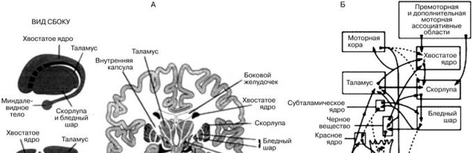
Subcortical (basal) nuclei refer to subcortical formations that have a common origin with the cerebral hemispheres and are located inside their white matter, between the frontal lobes and the diencephalon. These include caudate nucleus And shell, united by the common name "striated body" because the cluster nerve cells, forming gray matter, alternates with layers of white matter. Together with pale ball they form striopallidar system of subcortical nuclei. The striopallidary system also includes the claustrum, the subthalamic (subtubercular) nucleus, and the substantia nigra (Fig. 1).

Rice. 1. Basal nuclei of the brain and their connections with other systems: A - anatomy of the basal nuclei; B - connections of the basal nuclei with the corticospinal and cerebellar systems that control movement
The striopallidar system is the link between the cortex and the brain stem. Afferent and efferent pathways are suitable for this system.
Functionally, the basal nuclei are a superstructure over the red nuclei of the midbrain and provide plastic tone, i.e. the ability to hold for a long time an innate or learned pose, for example, the pose of a cat that guards a mouse, or a long holding of a pose by a ballerina performing some kind of step. When the cerebral cortex is removed, "wax rigidity" is observed, which is an expression of plastic tone without the regulatory influence of the cerebral cortex. An animal deprived of the cerebral cortex freezes for a long time in one position.
The subcortical nuclei ensure the implementation of slow, stereotyped, calculated movements, and the centers of the basal ganglia - the regulation of congenital and acquired movement programs, as well as the regulation of muscle tone.
Violation of various structures of the subcortical nuclei is accompanied by numerous motor and tonic shifts. So, in newborns, incomplete maturation of the basal ganglia leads to sharp convulsive flexion movements. As these structures develop, smoothness and calculated movements appear.
One of the main tasks of the basal ganglia in the implementation of motor control is the control of complex stereotypes of motor activity (for example, writing the letters of the alphabet). When there is severe damage to the basal ganglia, the cerebral cortex cannot properly maintain this complex stereotype. Instead, reproducing what has already been written becomes difficult, as if one had to learn to write for the first time. Examples of other stereotypes that are provided by the basal ganglia are cutting paper with scissors, hammering a nail, digging with a shovel in the ground, controlling eye and voice movements, and other well-practiced movements.
Caudate nucleus plays an important role in the conscious (cognitive) control of motor activity. Most of our motor acts arise as a result of their reflection and comparison with the information available in memory.
Violation of the functions of the caudate nucleus is accompanied by the development of hyperkinesis such as involuntary facial reactions, tremor, athetosis, chorea (twitching of the limbs, torso, as in an uncoordinated dance), motor hyperactivity in the form of aimless movement from place to place.
The caudate nucleus takes part in speech, motor acts. So, with a disorder of the anterior part of the caudate nucleus, speech is disturbed, difficulties arise in turning the head and eyes in the direction of sound, and damage to the posterior part of the caudate nucleus is accompanied by a loss vocabulary, decrease short term memory, cessation of voluntary breathing, speech delay.
Irritation striatum leads to sleep in the animal. This effect is explained by the fact that the striatum causes inhibition of the activating influences of the nonspecific nuclei of the thalamus on the cortex. The striatum regulates a number of vegetative functions: vascular reactions, metabolism, heat generation and heat release.
pale ball regulates complex motor acts. When it is irritated, a contraction of the muscles of the limbs is observed. Damage to the pale ball causes masking of the face, tremor of the head, limbs, monotony of speech, combined movements of the arms and legs when walking are disturbed.
With the participation of the pale ball, the regulation of orientation and defensive reflexes is carried out. When the pale ball is disturbed, food reactions change, for example, a rat refuses food. This is due to the loss of connection between the globus pallidus and the hypothalamus. In cats and rats, there is a complete disappearance of food-procuring reflexes after bilateral destruction of the globus pallidus.
- Subcortex ………………………………………………..page 8
- thalamus
- Hypothalamus
- Basal nuclei
- hippocampus
- amygdala
- midbrain
- Reticular formation
- Subcortical functions
- Conclusion
- List of used literature
The brain is a highly specialized part of the central nervous system. In humans, its mass averages 1375 g. It is here that huge clusters of intercalary neurons store the experience of actions gained throughout life. The brain is represented by 5 divisions. Three of them are the medulla oblongata, the pons and midbrain- are united under the name of the stem (or - the stem part) of the brain.
The stem part is fundamentally different from the other two parts of the brain, as it is supplied with cranial nerves, through which the stem directly controls the head and part of the neck. The other two departments - the diencephalon and the telencephalon - do not directly affect the structures of the human body, they regulate their functions, affect the centers of the trunk and spinal cord. The latter, equipped with cranial and spinal nerves, transmit through them generalized commands to performers - muscles and glands.
In addition to clusters of neurons that are directly related to nerves, the brain stem also contains other nerve centers that are similar in character to the centers of the diencephalon and telencephalon (reticular formation, red nuclei, black substance), which significantly distinguishes it from the spinal cord. A special place is occupied by the cerebellum, which performs the most important tasks of maintaining the degree of muscle tension (tonus), coordinating their work in performing movements, and maintaining balance at the same time. The cerebellum has a huge number of intercalary neurons, which it accommodates solely because they are not only in its thickness, but also in the extreme folded surface, making up the cerebellar cortex. Such a phenomenon manifests itself, in addition to it, only in the cortex of the telencephalon.
In front of and above the trunk is the diencephalon and the main components in the form of the visual tubercle (thalamus) - an important intermediate center along the sensory pathways to the telencephalon, hypothalamic region (hypothalamus) - it contains a lot of centers that are important for regulating metabolism in the body, its behavior and is closely related to the functioning of the pituitary gland, with which the leg is connected. Behind the visual hillock is the epiphysis (pineal gland) - an endocrine gland involved in the regulation of pigment metabolism in the skin and puberty.
The largest part of the mass of the brain is the telencephalon, usually described as two cerebral hemispheres connected by the corpus callosum. Its surface is sharply folded due to the mass of furrows (lateral, central, etc.) separating the gyri. Many of them are permanent, which makes it possible to distinguish areas of the cortex.
The hemispheres are divided into 4 main lobes. The frontal lobe is largely associated with the definition personal qualities of a person, and all the motor centers of the trunk and spinal cord are subordinate to its back. Therefore, when it is damaged, muscle paralysis appears. In the parietal lobe, sensations of heat, cold, touch, and the position of body parts in space are mainly formed. The occipital lobe contains visual centers, the temporal lobe - auditory and olfactory.
In the depths of the hemispheres, neurons are concentrated in the form of nodes (subcortex). They, together with other centers and the cerebellum, ensure the coordination of muscle work when performing motor programs of varying complexity. The brain is surrounded complex system shells. The soft shell is fused with its substance and contains the vessels that feed the brain, the branches of which penetrate into the thickness of the brain. Between it and the more superficial arachnoid, very thin and avascular, is the subarachnoid space with cerebrospinal fluid. Most of it is produced in the cavities of the brain (ventricles) and through the holes between the medulla oblongata and the cerebellum enters this space, forming a protective hydraulic cushion around the brain. Outermost hard shell connects to the bones of the skull.
SUBCORTICAL STRUCTURES OF THE BRAIN - parts of the brain located between the cerebral cortex and the medulla oblongata. They have an activating effect on the cortex, participate in the formation of all behavioral reactions in humans and animals, in maintaining muscle tone, etc.
Subcortical formations include structures located between the cerebral cortex and the medulla oblongata: the thalamus, hypothalamus, basal ganglia, a complex of formations combined into the limbic system of the brain, as well as the reticular formation of the brain stem and thalamus. Any afferent excitation arising from stimulation of receptors in the periphery is transformed into two streams of excitations at the level of the brainstem. One stream along specific paths reaches the projection area of the cortex, specific for a given irritation; the other one enters the reticular formation along the collaterals, from where it is directed to the cerebral cortex in the form of an ascending flow of excitations, activating it. The reticular formation has close functional and anatomical connections with the hypothalamus, thalamus, medulla oblongata, limbic system, cerebellum, so many activities of the body (breathing, food and pain reactions, motor acts, etc.) are carried out with its mandatory participation.
Afferent streams of excitations from peripheral receptors on their way to the cerebral cortex have numerous synaptic switches in the thalamus. From the lateral group of nuclei of the thalamus (specific nuclei), excitations are directed along two paths: to the subcortical ganglia and to specific projection zones of the cerebral cortex. The medial group of thalamic nuclei (nonspecific nuclei) serves as a switching point for ascending activating influences that are directed from the stem reticular formation to the cerebral cortex. Close functional relationships between specific and nonspecific nuclei of the thalamus provide the primary analysis and synthesis of all afferent excitations entering the brain. In animals at low stages of phylogenetic development, the thalamus and limbic formations play the role of the highest center of behavior integration, providing all the necessary motor acts of the animal aimed at preserving its life. In higher animals and humans, the highest center of integration is the cerebral cortex.
The limbic system includes a complex of brain structures that plays a leading role in the formation of the main innate reactions of humans and animals: food, sexual and defensive. It includes the lumbar gyrus, the hippocampus, the pyriform gyrus, the olfactory tubercle, the amygdala complex, and the septal region. The central place among the formations of the limbic system is given to the hippocampus. Anatomically, the hippocampal circle (hippocampus - fornix of the brain - mammillary bodies - anterior nuclei of the thalamus - cingulate gyrus - hippocampus) is established, which, together with the hypothalamus, plays a leading role in the formation of emotions. The regulatory influences of the limbic system extend widely to autonomic functions (maintenance of constancy internal environment body, regulation of blood pressure, respiration, vascular tone, motility of the gastrointestinal tract, sexual functions).
The cerebral cortex exerts constant downward (inhibitory and facilitating) influences on the subcortical structures. Exist various forms cyclic interaction between the cortex and subcortical structures, expressed in the circulation of excitations between them. The most pronounced closed cyclic connection exists between the thalamus and the somatosensory area of the cerebral cortex, which functionally constitute a single whole. The cortical-subcortical circulation of excitations can serve as the basis for the formation of the conditioned reflex activity of the organism.
Thalamus (thalamus, visual tubercle) is a structure in which the processing and integration of almost all signals going to the cerebral cortex from the spinal cord, midbrain, cerebellum, and basal ganglia of the brain takes place.
Morphofunctional organization. In the nuclei of the thalamus, the information coming from the extero-, proprioreceptors and interoceptors is switched and thalamocortical pathways begin.
Given that the geniculate bodies of the thalamus are the subcortical centers of vision and hearing, and the frenulum node and the anterior visual nucleus are involved in the analysis of olfactory signals, it can be argued that the thalamic thalamus as a whole is a subcortical “station” for all types of sensitivity. Here, the stimuli of the external and internal environment are integrated, after which they enter the cerebral cortex.
The visual hillock is the center of the organization and realization of instincts, drives, emotions. The ability to receive information about the state of many body systems allows the thalamus to participate in the regulation and determination of the functional state of the body as a whole (this is confirmed by the presence of about 120 multifunctional nuclei in the thalamus). The nuclei form peculiar complexes that can be divided according to the projection into the cortex into 3 groups: the anterior one projects the axons of its neurons into the cingulate gyrus of the cerebral cortex; medial - in the frontal lobe of the cortex; lateral - in the parietal, temporal, occipital lobes of the cortex. The function of the nuclei is also determined from the projections. Such a division is not absolute, since one part of the fibers from the nuclei of the thalamus goes to strictly limited cortical formations, the other to different areas of the cerebral cortex.
The nuclei of the thalamus are functionally divided into specific, nonspecific and associative, according to the nature of the incoming and outgoing pathways.
Specific nuclei include the anterior ventral, medial, ventrolateral, postlateral, postmedial, lateral, and medial geniculate bodies. The latter belong to the subcortical centers of vision and hearing, respectively.
The basic functional unit of specific thalamic nuclei are "relay" neurons, which have few dendrites and a long axon; their function is to switch information going to the cerebral cortex from skin, muscle and other receptors.
From specific nuclei, information about the nature of sensory stimuli enters strictly defined areas of III-IV layers of the cerebral cortex (somatotopic localization). Violation of the function of specific nuclei leads to the loss of specific types of sensitivity, since the nuclei of the thalamus, like the cerebral cortex, have somatotopic localization. Individual neurons of specific nuclei of the thalamus are excited by receptors of only their own type. Signals from the receptors of the skin, eyes, ear, and muscular system go to the specific nuclei of the thalamus. Signals from the interoreceptors of the projection zones of the vagus and celiac nerves, the hypothalamus also converge here.
The lateral geniculate body has direct efferent connections with the occipital lobe of the cerebral cortex and afferent connections with the retina and anterior colliculi. The neurons of the lateral geniculate bodies react differently to color stimuli, turning on and off the light, i.e., they can perform a detector function.
The medial geniculate body (MC) receives afferent impulses from the lateral loop and from the inferior tubercles of the quadrigeminae. The efferent pathways from the medial geniculate bodies go to the temporal zone of the cerebral cortex, reaching there the primary auditory cortex. MKT has a clear tonotopicity. Consequently, already at the level of the thalamus, the spatial distribution of the sensitivity of all sensory systems of the body, including sensory transmissions from the interoreceptors of blood vessels, organs of the abdominal and chest cavities, is ensured.
The associative nuclei of the thalamus are represented by the anterior mediodorsal, lateral dorsal, and pillow nuclei. The anterior nucleus is associated with the limbic cortex (cingulate gyrus), the mediodorsal - with the frontal lobe of the cortex, the lateral dorsal - with the parietal, the pillow - with the associative zones of the parietal and temporal lobes of the cerebral cortex.
The main cellular structures of these nuclei are multipolar, bipolar three-pronged neurons, i.e., neurons capable of performing polysensory functions. A number of neurons change activity only with simultaneous complex stimulation. Convergence of excitations of different modalities occurs on polysensory neurons, an integrated signal is formed, which is then transmitted to the associative cortex of the brain. The neurons of the pillow are mainly associated with the associative zones of the parietal and temporal lobes of the cerebral cortex, the neurons of the lateral nucleus - with the parietal, the neurons of the medial nucleus - with the frontal lobe of the cerebral cortex.
Nonspecific nuclei of the thalamus are represented by the median center, paracentral nucleus, central medial and lateral, submedial, ventral anterior, parafascicular complexes, reticular nucleus, periventricular and central gray mass. The neurons of these nuclei form their connections according to the reticular type. Their axons rise to the cerebral cortex and contact with all its layers, forming not local, but diffuse connections. Connections from the RF of the brain stem, hypothalamus, limbic system, basal ganglia, and specific nuclei of the thalamus come to nonspecific nuclei.
Excitation of nonspecific nuclei causes the generation of specific spindle-shaped electrical activity in the cortex, indicating the development of a sleepy state. Violation of the function of nonspecific nuclei makes it difficult for spindle-shaped activity to appear, i.e., the development of a sleepy state.
Pages:123456next →
Projection fibers are those fibers that connect the hemispheres of the brain with the underlying parts of the brain - the stem and spinal cord. As part of the projection fibers, there are conducting paths that carry afferent (sensitive) and efferent (motor) information.
So, the main sections, furrows and convolutions of the brain are shown in Fig. 5.6.
Rice. 5. Brain, left hemisphere (side view):
1 - precentral gyrus; 2 - precentral furrow; 3 - superior frontal gyrus; 4 - central furrow; 5 - middle frontal gyrus;
b - inferior frontal gyrus; 7 - ascending branch of the lateral furrow;
8 - horizontal branch of the lateral furrow; 9 - posterior branch of the lateral furrow; 10 - superior temporal gyrus; 11 - middle temporal gyrus;
12 - inferior temporal gyrus; 13 - parietal lobule; 14 - post-central furrow; 15 - postcentral gyrus; 16 - supramarginal gyrus;
17 - angular gyrus; 18 - occipital lobe; 19 - cerebellum; 20 - horizontal fissure of the cerebellum; 21 - medulla oblongata
The structure of the subcortical region of the brain. diencephalon
2.1 Striopallidar system
In the thickness of the white matter of the cerebral hemispheres there are accumulations of gray matter called the subcortical nuclei (basal nuclei). These include the caudate nucleus, the lenticular nucleus, the fence and the amygdala (Fig. 6). The lenticular nucleus, located outside the caudate nucleus, is divided into three parts. It distinguishes between the shell and two pale balls.

Rice. 6. Subcortical nuclei:
1 - caudate nucleus; 2 - lenticular nucleus; 3 - visual tubercle.
A - horizontal section: a - fence; b - shell; c and d - pale ball;
B-frontal section: a - pale ball; b - shell
In functional terms, the caudate nucleus and the shell are combined into the striatum (striatum), and the pale balls, together with the black substance and red nuclei located in the legs of the brain, into the pale body (pallidum).
Together they represent a very functionally important formation - the strioppallidar system. According to morphological features and phylogenetic origin (their appearance at a certain stage of evolutionary development), the pale body is more ancient than the striatum formation.
Basal structures
The striopallidary system is important integral part propulsion system. It is part of the so-called pyramid system. In the motor zone of the cerebral cortex, the motor - pyramidal - path begins, along which the order follows to perform a particular movement.
The extrapyramidal system, an important part of which is the striopallidum, being included in the motor pyramidal system, takes an auxiliary part in providing voluntary movements.
At a time when the cerebral cortex was not yet developed, the striopallidar system was the main motor center that determined the behavior of the animal. Due to the striopallidar locomotor apparatus, diffuse, mass, body movements were carried out, providing movement, swimming, etc.
With the development of the cerebral cortex, the striopallidary system passed into a subordinate state. The main motor center was the cerebral cortex.
The striopallidary system began to provide a background, a readiness to make a movement; against this background, fast, precise, strictly differentiated movements controlled by the cerebral cortex are carried out.
To make a movement, it is necessary that some muscles contract and others relax, in other words, you need an accurate and coordinated redistribution of muscle tone.
Such a redistribution of muscle tone is precisely carried out by the striopallidar system. This system provides the most economical consumption of muscle energy in the process of performing a movement. Improving the movement in the process of learning to perform them (for example, working to the limit of a musician’s honed finger run, a mower’s wave of a hand, precise movements of a car driver) leads to gradual economization and automation.
This possibility is provided by the striopallidar system.
It was noted above that, phylogenetically, the striatum is a younger formation than the pale body. Fish are an example of pallidar organisms.
They move in the water with the help of throwing powerful movements of the body, without "caring" about saving muscle energy. These movements are relatively precise and powerful. However, they are energetically wasteful. In birds, the striatum is already well defined, which helps them more prudently regulate the quality, accuracy and quantity of movements. Thus, the pale body inhibits and regulates the activity of the pallidar system (i.e.,
phylogenetically younger formations control and inhibit older ones).
The motor acts of the newborn are pallidary in nature: they are uncoordinated, throwing and often superfluous. With age, as the striatum matures, the child's movements become more economical, stingy, automated.
The striopallidar system has connections with the cerebral cortex, cortical motor system (pyramidal) and muscles, formations of the extrapyramidal system, with the spinal cord and thalamus.
Other basal nuclei (fence and amygdala) are located outward from the lentiform nucleus. The amygdala enters another functional system- limbic-reticular complex.
2.2 thalamus
The optic tubercle and the hypothalamus (hypothalamus) develop from the intermediate cerebral bladder, and the third ventricle develops from the cavity of the intermediate cerebral bladder.
visual bulge, or thalamus, located on the sides of the third ventricle and consists of a powerful accumulation of gray matter.
The visual tubercle is divided into the actual visual tubercle, the epithalamic (suprathalamic region, or epilamus) and the foreign (zathalamic region, or metathalus). The bulk of the gray tubercle is the thalamus (see Fig. 7).

Rice. 7. Topography of the thalamus
1 - thalamus; 2 - body of the caudate nucleus; 3 - body of the lateral ventricle; 4 - corpus callosum; 5 - medulla oblongata.
A protrusion of a pillow is distinguished in it, posterior to which there are two elevations - the outer and inner cranked bodies (they enter the foreign region).
There are several nuclear groups in the thalamus.
The epithalamus, or epithalamus, consists of the pineal gland and the posterior commissure of the brain.
The foreign region, or metathalamus, includes the geniculate bodies, which are the elevation of the thalamus. They lie outward and downward from the pillow of the thalamus.
The hypothalamic region, or hypothalamus, lies down from the thalamus, has a number of nuclei lying in the walls of the third ventricle.
The visual tubercle is an important stage on the way to conduct all types of sensitivity. Sensory pathways are approached and concentrated in it - touch, pain, temperature sensation, visual tracts, auditory pathways, olfactory pathways and fibers from the extrapyramidal system. From the neurons of the thalamus begins the next stage of transmission of sensitive impulses - to the cerebral cortex.
At a certain stage in the evolution of the nervous system, the thalamus was the center of sensitivity, just as the Striopallidar system was the mechanism of movements. As the cerebral cortex appeared and developed, the main role in the function of the sensitive sphere was transferred to the cerebral cortex, and the visual tubercle remained only a transmission station for sensitive impulses from the periphery to the cerebral cortex.
Midbrain, quadrigemina. Structure and functions. Sylvius aqueduct. Stem part of the brain
Midbrain, quadrigemina
In the roof of the midbrain, a plate in the form of a quadrigemina is distinguished. The two upper hillocks, as already mentioned above, are the subcortical centers of the visual analyzer, and the lower ones are the auditory analyzer.
The quadrigemina is a reflex center of various kinds of movements that arise mainly under the influence of visual and auditory stimuli. It is here that the impulses are switched to the underlying structures of the brain.
The roof of the midbrain (tectum mesencephali) is a plate of the quadrigemina (lamina quadrigemina), which includes two pairs of tubercles (mounds): the upper tubercles (colliculi superiores, mounds) of the quadrigemina and the lower tubercles (colliculi inferiores, mounds) of the quadrigemina.
The upper tubercles (knolls) in humans are slightly larger than the lower ones.
Between the upper tubercles there is a wide depression, which is called the subpineal triangle. Above this cavity is the epiphysis ( pineal gland) .
From each hillock in the lateral direction, a thickening in the form of a roller, which is a bundle of fibers, departs. These are the handle of the upper mound (brachium colliculi cranialis, superiors) and the handle of the lower mound (brachium colliculi caudalis, inferioris).
The handles of the hillocks go to the diencephalon.
The handle of the superior colliculus runs under the thalamic cushion to the lateral geniculate body and to the optic tract. The wider and flatter lower handle disappears under the medial geniculate body. The thalamic cushion, geniculate bodies, and optic tract already belong to the diencephalon.
Subcortical regions of the brain (subcortex)
In humans, the superior colliculus of the roof of the midbrain and the lateral geniculate bodies function as midbrain visual centers, and the inferior colliculus of the quadrigemina and medial geniculate bodies act as auditory centers.
Sylvius aqueduct
Inside the midbrain there is a narrow canal - the aqueduct of the brain (Sylvian aqueduct).
The aqueduct of Sylvius is a narrow canal 2 cm long that connects the III and IV ventricles. Around the aqueduct is the central gray matter, which contains the reticular formation, nuclei III and nuclei IV of pairs of cranial nerves, etc.
On sections of the midbrain, the Sylvian aqueduct may look like a triangle, rhombus, or ellipse.
The aqueduct of the brain connects the third ventricle of the diencephalon and the fourth ventricle of the rhomboid brain. Around the aqueduct of the midbrain is the central gray matter (substantia grisea centralis). In the central gray matter in the region of the bottom of the aqueduct lie the nuclei of two pairs of cranial cerebral nerves: at the level of the upper colliculus of the quadrigemina is the nucleus of the oculomotor nerve (III pair); at the level of the lower mounds of the quadrigemina lies the nucleus of the trochlear nerve (IV pair).
Previous12131415161718192021222324252627Next
On the influence of the cortex on the cortex through subcortical formations.
It was also shown in some old works that the same diffuse inhibition of cortical rhythms, which is obtained by stimulation of the reticular formation, can also occur with stimulation of the cortex (Dusser de Barenne, McCulloch, 27; Beritov, Bregadze, Tskipuridze, 28). Later, the influence of stimulation of the limbic cortex (orbital cortex and anterior cingulate gyrus) on the slow electrical activity of other cortical regions in cats and monkeys was studied in detail (Sloan and Jasper, 29).
Immediately after the cessation of short-term stimulation of the cingulate gyrus in different parts of the brain, a significant change in electrical activity occurred, which lasted a relatively long time, gradually returning to its original state.
The effect was generalized, i.e., changes were observed both on the surface of the cortex of both hemispheres and in the thalamic specific and nonspecific nuclei.
In the overwhelming majority of cases, a decrease in the amplitude of slow potentials was obtained, up to their complete suppression.
It is important to note that the generalized nature of the effect of irritation of the limbic cortex was not obtained due to transcortical distribution
injury of excitation, but through the activation of a subcortical mechanism that acts diffusely on the cortex, i.e.
e. through corticofugal excitation of nonspecific formations of the thalamus and trunk. This was evident from the fact that after the subpial incision and isolation of the irritated area from the rest of the cortex, the effect was not eliminated. After trimming the white matter under the irritated area of the cortex, the effect disappeared. Almost the same results were also obtained by Kaada when stimulating the anterior "rhinencephalic" areas of the cortex (Kaada, 30).
Meaning new cortex in the regulation of the function of the ascending reticular system has been systematically and in detail studied throughout recent years Bremer with employees.
Thus, Bremer and Terzuolo (31) showed on isolated encephalic preparations of a cat the important role of the cerebral cortex in awakening and maintaining the waking state. This was evident from the fact that after bilateral coagulation of the primary and secondary auditory cortex zones to semantic sound stimuli (call), the dormant cat preparation no longer awakens (judging by the electrical activity of the cortex and eye reactions), while skin stimuli retain their awakening power.
These experiments directly indicate that awakening in response to sound stimuli, which have the value of a conditioned signal, can be realized only through the primary excitation of a specific auditory area of the cortex, which, acting on the reticular formation, excites it, and this latter already activates the entire cortex for the second time and causes awakening.
It is clear that when a cat calls, excitation impulses from the receptor are directed in the same way as with weak sound stimulation: both to the corresponding perceiving area of the cortex and to the reticular formation. When both points receiving sound impulses function normally, in other words, when there is a normal interaction between the cortex and the reticular formation and, in particular, when the downward influence of the cortex on the reticular formation is not disturbed, then the call of the animal awakens.
But if the cortical auditory areas are turned off, then the same afferent impulses coming to the reticular formation during the call are no longer able to cause awakening.
The role of corticofugal impulses in awakening is also indicated by the experiments of Roitbak and Buthuzi (32). They were able to observe the awakening response (both behavioral and EEG) in response to direct electrical stimulation of the internal geniculate body in cats with chronically implanted electrodes.
It is also possible that this awakening is the result of primary excitation of the auditory cortex, which in turn activates the reticular formation and thereby causes the animal to awaken.
In the experiments of Bremer and Terzuolo (31) with direct electrical stimulation of different parts of the neocortex, response potentials of the same nature were registered in the reticular formation, which, when following one after another, like the responses of peripheral stimuli, acted both facilitating and depressing.
Mutual influence in the reticular formation was also exerted by corticofugal and peripheral impulses. Finally, it was shown that short-term tetanic stimulation of different parts of the cortex causes exactly the same pattern of awakening (bilateral "desynchronization" of the EEG, pupil dilation and eye movements) that is observed during activation of the reticular formation caused by its direct electrical stimulation or through peripheral afferent impulses.
The bilateral "awakening reaction" of the EEG does not disappear after sectioning the corpus callosum; it occurs during the period of cortical irritation and lasts long after the stimulation has ceased. Of the tested areas of the cortex, the most effective were the somatosensory, somatomotor and parastriate areas.
Irritation of the cortex affects both the reticular formation and other subcortical formations. This has been studied in detail in monkeys (French, Hernandez-Peon and Livingston, 33).
First of all, it was shown that
regardless of the irritated area of the cortex, response potentials occur simultaneously in a wide area of the subcortex, starting from the anterior commissure to the pontine tegmentum. Along with the reticular formation and nonspecific thalamic nuclei, responses to cortical irritation were recorded in the caudate nucleus, the globus pallidus, the substantia nigra, the red nucleus, the thalamic transmission and association nuclei, etc.
e. Thus, each of the irritated areas of the cortex turned out to be connected by descending pathways with a wide area of the brain stem, and each area of one or another subcortical formation - with almost all areas of the cortex. Due to this, when stimuli were combined in two different parts of the cortex, their responses in the reticular formation mutually influenced each other.
In the same areas of the subcortex, responses to irritation of the sciatic nerve were also recorded. The response potentials of the trunk to stimulation of both the cortex and the sciatic nerve were equally inhibited after the administration of barbiturate. In local strychnine poisoning of the cortex, synchronously with cortical strychnine discharges, discharges or waves of the same nature appeared in nonspecific thalamic nuclei and the reticular formation, or their background activity changed markedly.
However, it has been shown that not all areas of the cortex have the same effect on the subcortex.
The sensorimotor, posterior parietal or paraoccipital regions, superior temporal gyrus, orbital surface of the frontal lobe and cingulate gyrus turned out to be the most effective, while irritation of the frontal and occipital poles and the baeal surface temporal lobe did not give answers.
In terms of their effect on the trunk, individual areas of the cortex did not differ from each other in any way: they elicited the same responses with approximately the same latent period in all the studied subcortical formations.
Subcortical functions
EEG desynchronization is considered an essential electrical correlate of behavioral arousal, and therefore it can be concluded that stimulation of the above cortical areas can also cause behavioral arousal.
This phenomenon was specifically studied in monkeys with implanted electrodes. The electrodes were implanted in different areas of the cortex, including those areas whose irritation caused the appearance of response potentials in the reticular formation. It has been shown that a sleeping animal is quickly awakened by stimulation if it is produced in the aforementioned areas of the cortex (sensomotor, posterior parietal, superior temporal, orbital and cingulate gyrus).
Irritation of other areas of the cortex did not cause awakening. If the active cortical areas were irritated in the waking state, then a pattern developed indicating the animal's alertness (immediate cessation of movements, freezing in one position, raising the head and turning into different sides etc.). In other words, a typical orienting reaction developed from irritation of the reticular formation itself.
It is well known that during wakefulness, drowsiness or sleep in an unanesthetized animal (with implanted electrodes) in the cortex and many subcortical formations (including in the reticular formation of the trunk), in most cases, the same activity is recorded; for example, during sleep, both in the cortex and in the subcortical formations, slow activity occurs almost simultaneously (Narikashvili, 2; Tskipuridze, 5; Bremer and Tertsuolo, 31).
The same was observed by Jouvet and Michel (34) in cats in which the electrodes were implanted in different areas of the cortex and the mesodiencephalic region. However, after mesencephalic transection or after removal (or damage) of the entire cortex (by suction or coagulation), during natural or barbiturous sleep of the animal in the subcortical formations (with the exception of the hippocampus),
thorny for a dream slow activity did not develop any more.
For the development of slow activity in the reticular formations, it was necessary to maintain, in connection with the latter, a certain minimum mass of the neocortical substance. They failed to establish any predominant role of certain areas of the cortex. Thus, that restructuring of the activity of neurons in the reticular formations, which leads to their synchronous activity and the appearance of slow waves, does not occur without connection with the cortex (see also Serkov, et al., 35).
It turns out that the cortex is directly involved in the organization of the electrical rhythms of reticular formations, just as the latter are directly involved in the formation and change of cortical rhythms. This once again confirms the idea of a close connection and unity of the function of these formations.
Summarizing all of the above, we can conclude that the reticular ascending activating system can be excited by both corticofugal and peripheral afferent impulses.
Both different corticofugal and different peripheral impulses converge to the same areas or neurons of the reticular formation, due to which an interaction is observed both between corticofugal impulses coming from different areas of the cortex, and between them and peripheral impulses. "Activation" or "desynchronization" of the EEG, as well as the awakening of the animal, can occur both from peripheral and from corticofugal impulses.
Previous67686970717273747576777879808182Next
VIEW MORE:
provide regulation of vital processes in the body due to the activity of subcortical formations of the brain. The subcortical structures of the brain have functional differences from the cortical structures and occupy a conditionally subordinate position in relation to the cortex. The basal ganglia, thalamus, and hypothalamus were first referred to as such structures. Later, the striopallidar system (see Extrapyramidal system) was identified as physiologically independent systems, including the basal ganglia and midbrain nuclear formations (red nucleus and black substance); thalamocortical system: reticulocortical system (see.
Reticular formation), limbic-neocortical system (see. Limbic system), cerebellar system (see. Cerebellum), system of nuclear formations of the diencephalon, etc.
(rice.).
Subcortical functions play an important role in the processing of information entering the brain from external environment and the internal environment of the body. This process is provided by the activity of the subcortical centers of vision and hearing (lateral, medial, geniculate bodies), primary centers for the processing of tactile, pain, protopathic, temperature and other types of sensitivity - specific and nonspecific nuclei of the thalamus.
A special place among P.
Subcortical structures of the brain
f. occupy the regulation of sleep (Sleep) and wakefulness, the activity of the hypothalamic-pituitary system (Hypothalamo-pituitary system), which ensures the normal physiological state of the body, homeostasis. An important role belongs to P. f. in the manifestation of the main biological motivations of the body, such as food, sex (see Motivation).P. f. are realized by emotionally colored forms of behavior; of great clinical and physiological importance are P. t. in the mechanisms of manifestation of convulsive (epileptiform) reactions of various origins.
Thus, P. f. are the physiological basis of the activity of the entire brain. In turn, P. f. are under constant modulation higher levels cortical integration and mental sphere.
With lesions of the subcortical structures, the clinical picture is determined by the localization and nature of the pathological process.
For example, damage to the basal ganglia is usually manifested by Parkinson's syndrome, extrapyramidal hyperkinesis (Hyperkinesis). The defeat of the nuclei of the thalamus is accompanied by disorders of various types of sensitivity (Sensitivity), movements (Movement), regulation of autonomic functions (see Vegetative nervous system). Disturbances in the function of deep structures (brain stem, etc.) manifest themselves in the form of bulbar paralysis (bulbar paralysis), pseudobulbar paralysis (pseudobulbar paralysis) with a severe outcome.
See also Brain, Spinal cord.

Schematic representation of the main afferent and efferent connections in the process of implementing subcortical functions.
Parts of the brain located between the cerebral cortex and the medulla oblongata. They have an activating effect on the cortex, participate in the formation of all behavioral reactions of humans and animals, in maintaining muscle tone, etc. Big encyclopedic Dictionary
Parts of the brain located between the cerebral cortex and the medulla oblongata. They have an activating effect on the cortex, participate in the formation of all behavioral reactions of humans and animals, in maintaining muscle tone, etc. * * ... ... encyclopedic Dictionary
Parts of the brain located between the cerebral cortex and the medulla oblongata They have an activating effect on the cortex, participate in the formation of all behavioral reactions in humans and animals, in maintaining muscle tone, etc ... Natural science. encyclopedic Dictionary
subcortex (subcortical structures of the brain) The part of the brain located between the cerebral cortex and the medulla oblongata. It includes: visual tubercles, hypothalamus, limbic system and other basal nerve nodes, reticular formation of the brain stem, thalamus. P. participates ... Encyclopedic Dictionary of Psychology and Pedagogy
The totality of physiological processes associated with the activity of individual subcortical structures of the brain (See Subcortical structures of the brain) or with their system. From an anatomical point of view, all ganglion formations are classified as subcortical, ... ...
SUBCORTICAL FUNCTIONS- SUBCORTAL FUNCTIONS. The doctrine of the functions of P. formations, which developed on the basis of anat. clinical (predominantly) comparatively anatomical and experimental physiological studies, is i.e many years old and cannot be considered as ... Big Medical Encyclopedia
A layer of gray matter 1 5 mm thick covering the cerebral hemispheres of mammals and humans. This part of the brain (See Brain), which developed in the later stages of the evolution of the animal world, plays exclusively ... ... Great Soviet Encyclopedia
100 r first order bonus
Choose the type of work Graduate work Course work Abstract Master's thesis Report on practice Article Report Review Test Monograph Problem solving Business plan Answers to questions creative work Essay Drawing Compositions Translation Presentations Typing Other Increasing the uniqueness of the text Candidate's thesis Laboratory work Help online
Ask for a price
The forebrain consists of subcortical (basal) nuclei and the cerebral cortex. The subcortical nuclei are part of the gray matter of the cerebral hemispheres and consist of the striatum, globus pallidus, shell, fence, subthalamic nucleus and substantia nigra. The subcortical nuclei are the link between the cortex and the brain stem. Afferent and efferent pathways approach the basal ganglia.
Functionally, the basal nuclei are a superstructure over the red nuclei of the midbrain and provide plastic tone, i.e. the ability to hold a congenital or learned posture for a long time. For example, the pose of a cat that guards a mouse, or a long-term retention of a pose by a ballerina performing some kind of step.
The subcortical nuclei allow for slow, stereotyped, calculated movements, and their centers - the regulation of muscle tone.
Violation of various structures of the subcortical nuclei is accompanied by numerous motor and tonic shifts. So, in a newborn, incomplete maturation of the basal nuclei (especially the pale ball) leads to sharp convulsive flexion movements.
Violation of the function of the striatum leads to a disease - chorea, accompanied by involuntary movements, significant changes in posture. With a disorder of the striatum, speech is disturbed, there are difficulties in turning the head and eyes towards the sound, there is a loss of vocabulary, and voluntary breathing stops.
Subcortical functions play an important role in the processing of information that enters the brain from the external environment and the internal environment of the body. This process is provided by the activity of the subcortical centers of vision and hearing (lateral, medial, geniculate bodies), primary centers for the processing of tactile, pain, protopathic, temperature and other types of sensitivity - specific and nonspecific nuclei of the thalamus. A special place among P. f. occupied by the regulation of sleep and wakefulness, the activity of the hypothalamic-pituitary system, which ensures the normal physiological state of the body, homeostasis. An important role belongs to P. f. in the manifestation of the main biological motivations of the body, such as food, sex. P. f. are realized by emotionally colored forms of behavior; of great clinical and physiological importance are P. t. in the mechanisms of manifestation of convulsive (epileptiform) reactions of various origins. Thus, P. f. are the physiological basis of the activity of the entire brain. In turn, P. f. are under the constant modulating influence of the higher levels of cortical integration and the mental sphere.
The basal ganglia develop faster than the optic thalamus. Myelination of the BU structures begins in the embryonic period, and ends by the first year of life. Physical activity newborn depends on the functioning of the globus pallidus. Impulses from it cause general uncoordinated movements of the head, torso, limbs. In the newborn, AEs are associated with the optic tubercles, hypothalamus, and substantia nigra. With the development of the striatum, the child develops facial movements, and then the ability to sit and stand. At 10 months, the baby can stand freely. As the basal ganglia and cerebral cortex develop, movements become more coordinated. By the end preschool age the balance of cortical-subcortical motor mechanisms is established.